MIDLMedical Imaging with Deep Learning
\jmlrpages
\jmlryear2019
\jmlrworkshopMIDL 2019 – Extended Abstract Track
\midlauthor\NameThibault Févry\nametag1, \NameJason Phang\nametag1, \NameNan Wu \nametag1, \NameS. Gene Kim\nametag2, \NameLinda Moy\nametag2,
\NameKyunghyun Cho\nametag1, \NameKrzysztof J. Geras\nametag2,1
\addr1 Center for Data Science, New York University
\addr2 Department of Radiology, New York University School of Medicine
Improving localization-based approaches for breast cancer screening exam classification
Abstract
We trained and evaluated a localization-based deep CNN for breast cancer screening exam classification on over 200,000 exams (over 1,000,000 images). Our model achieves an AUC of 0.919 in predicting malignancy in patients undergoing breast cancer screening, reducing the error rate of the baseline [Wu et al. (2019a)] by 23%. In addition, the models generates bounding boxes for benign and malignant findings, providing interpretable predictions.
1 Introduction
Breast cancer is the second leading cause of cancer-related death among women in the United States. Screening mammography has been highly effective in reducing mortality rate but suffers from a high rate of false positive recalls, leading to added costs and stress. Better computer-aided diagnostic tools could improve patient outcomes by helping radiologists. Our goal is to build a system that provides highly accurate as well as interpretable predictions, in order to assist radiologists in cancer detection. To this end, we train an object detection network to predict the location of suspicious lesions and classify them.
2 Data
Our dataset [Wu et al. (2019b)] comprises 229,426 screening mammography exams (1,001,093 images, with each exam containing the four standard views) from 141,473 unique patients. Among these, 5,832 exams had at least one biopsy performed within 120 days of a screening mammogram. Within this set of exams, 985 breasts had malignant findings, 5,556 had benign findings and 234 had both. For these exams, radiologists retrospectively annotated the locations of the biopsied lesions at a pixel level.
3 Methods
We used bounding boxes on the annotations of biopsied lesions to train a Feature-Pyramidal Network [Lin et al. (2017)]-based Faster-RCNN model [Ren et al. (2015)]. In most experiments, we used a ResNet-50 (R-50; He et al., 2016) backbone pretrained on ImageNet Deng et al. (2009) to initialize our models. We also conducted experiments with ResNet-101 (R-101) and ResNeXt-101 (X-101; Xie et al., 2017) backbones. The final classifier predicts three classes: benign lesions, malignant lesions, or nothing. We used maskrcnn-benchmark Massa and Girshick (2018) to train our models. We evaluated models after every 5,000 mini-batches and select the best models according to the score computed on the validation set. We used the standard setup of the framework, aside from the following exceptions.
Use of non-annotated images
We used images without annotated lesions to train our model. These are treated as negatives for both the region-proposal network (RPN) and the classifier branch. The goal of this approach is to reduce overfitting on the comparatively small set of annotations. For our base experiment, during training, half of the images are sampled from an exam with a biopsy and half without.
Resolution
We used a maximum resolution of for the R-50 backbone, by resizing the mammograms with bilinear interpolation ( for R-101, for X-101). Indeed, prior work Geras et al. (2017) highlighted the importance of operating at a high resolution. Consequently, we also reduced the batch size to 4 (2 per GPU) due to our model’s large memory requirements.
IoU thresholds
We also relaxed the IoU threshold for foreground objects in the RPN from 0.7 to 0.5, as in Ribli et al. (2018); Morrell et al. (2018). Compared to natural images, our data set has (i) noisier annotations with less precise boundaries, and (ii) fewer annotations per image. To circumvent (i), we also tried isotropic bounding box rescaling by a factor drawn uniformly at random in . We set the IoU threshold of the final non-maximum suppression to 0.1, following the rationale of Ribli et al. (2018) that for mammograms, “overlapping detections are expected to happen less often than in usual object detection".
Optimization
We generally did not tune the learning rate schedules according to the advice of Goyal et al. (2017) (“recommended lr schedule”) despite lowering the batch size. Indeed, found that it did not strongly impact results while considerably increasing training time. We used gradient clipping (for norm ) in all our experiments. We experimented with using batch normalization but found it not to help in our experiments.
Inference
During inference, we take the maximum malignant prediction among the bounding boxes of each view and then average over both views to get the predicted probability for the whole breast. We found this setup to perform better than others, including taking the maximum over views or the mean over boxes. In addition, we found it useful to decrease the score threshold at inference time from 0.05 to 0.001, as the tail-like behavior is important when computing AUC.
4 Results
| model | test set | reader study | ||
| Wu et al. (2019a) single model | 0.886 | 0.003 | - | |
| Wu et al. (2019a) ensemble | 0.895 | 0.876 | ||
| Base setup | 0.891 | 0.005 | 0.845 | 0.007 |
| + biopsy ratio 0.75 | 0.895 | 0.004 | 0.855 | 0.012 |
| + biopsy ratio 1 | 0.887 | 0.011 | 0.855 | 0.013 |
| + bounding box scaling | 0.890 | 0.006 | 0.841 | 0.010 |
| + classifier*5 | 0.903 | 0.007 | 0.859 | 0.013 |
| + bb scaling, classifier*5, ratio 0.75 | 0.888 | 0.006 | 0.855 | 0.006 |
| + recommended lr schedule | 0.897 | 0.007 | 0.858 | 0.007 |
| + R-101 backbone | 0.887 | 0.011 | 0.834 | 0.011 |
| + X-101 backbone | 0.908 | 0.014 | 0.866 | 0.020 |
| Ensemble | 0.919 | 0.879 | ||
| Ensemble + Wu et al. (2019a) ensemble | 0.930 | 0.895 | ||
The results are shown in \autoreftab:main_results. Our methods compare favorably against the models of Wu et al. (2019a). Indeed, we achieve a 23% relative error reduction in AUC on the test set. Note that both models use pixel-level annotations. Due to high variance, we ensemble all runs together for our "Ensemble" setup. On the subpopulation of the test set used for the reader study in Wu et al. (2019a), our results also compare favorably to both earlier models and radiologists in terms of ROC, although the improvements are smaller. The reader study setup contains more exams from patients who underwent biopsy. Thus we believe this model distinguishes better between negative cases and cases that need a biopsy but is not as good in distinguishing between benign and malignant cases within the biopsied population. Ensembling our method with Wu et al. (2019a) improves the reader study AUC further to 0.895, compared to 0.778 for the average radiologist.
5 Analysis
 |
 |
Our validation metrics varied significantly between checkpoints. We believe that this is due to (i) the interaction of the components of the training loss, (ii) components of the training loss being only loosely related to the final metric, (iii) a small batch size rendering optimization unstable. This motivates ensembling all runs to produce our final model.
We notice a clear trade-off between using larger networks and using a larger resolution, with the R-50, R-101 and X-101 setups performing competitively. Unfortunately, due to computational constraints, we were unable to test R-101 and X-101 on the same resolution as R-50. Overall, our ensemble combining different backbones and resolutions performed best.
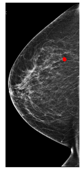
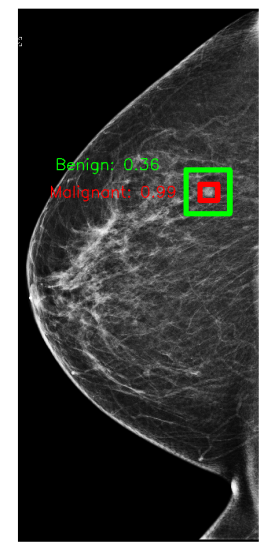
In Figure 1, we show an annotation and the corresponding prediction using an X-101 model. The model accurately predicts a malignant lesion with high probability (0.99). It also predicts a benign lesion with low probability (0.36) for which there is no ground-truth annotation. These bounding boxes can highlight suspicious regions and help radiologists understand predictions from our models.
We would like to thank Yiqiu Shen and Jungkyu Park for comments on earlier versions of this manuscript. We also gratefully acknowledge the support of Nvidia Corporation with the donation of some of the GPUs used in this research. This work was supported in part by grants from the National Institutes of Health (R21CA225175 and P41EB017183).
References
- Deng et al. (2009) J. Deng, W. Dong, R. Socher, L.-J. Li, K. Li, and L. Fei-Fei. ImageNet: A Large-Scale Hierarchical Image Database. In CVPR, 2009.
- Geras et al. (2017) Krzysztof J Geras, Stacey Wolfson, Yiqiu Shen, S Kim, Linda Moy, and Kyunghyun Cho. High-resolution breast cancer screening with multi-view deep convolutional neural networks. arXiv:1703.07047, 2017.
- Goyal et al. (2017) Priya Goyal, Piotr Dollár, Ross Girshick, Pieter Noordhuis, Lukasz Wesolowski, Aapo Kyrola, Andrew Tulloch, Yangqing Jia, and Kaiming He. Accurate, large minibatch sgd: Training imagenet in 1 hour. arXiv:1706.02677, 2017.
- He et al. (2016) Kaiming He, Xiangyu Zhang, Shaoqing Ren, and Jian Sun. Deep residual learning for image recognition. In CVPR, 2016.
- Lin et al. (2017) Tsung-Yi Lin, Piotr Dollár, Ross Girshick, Kaiming He, Bharath Hariharan, and Serge Belongie. Feature pyramid networks for object detection. In CVPR, 2017.
- Massa and Girshick (2018) Francisco Massa and Ross Girshick. maskrcnn-benchmark: Fast, modular reference implementation of Instance Segmentation and Object Detection algorithms in PyTorch. https://github.com/facebookresearch/maskrcnn-benchmark, 2018.
- Morrell et al. (2018) Stephen Morrell, Zbigniew Wojna, Can Son Khoo, Sebastien Ourselin, and Juan Eugenio Iglesias. Large-scale mammography cad with deformable conv-nets. In Image Analysis for Moving Organ, Breast, and Thoracic Images. 2018.
- Ren et al. (2015) Shaoqing Ren, Kaiming He, Ross Girshick, and Jian Sun. Faster r-cnn: Towards real-time object detection with region proposal networks. In NIPS, 2015.
- Ribli et al. (2018) Dezső Ribli, Anna Horváth, Zsuzsa Unger, Péter Pollner, and István Csabai. Detecting and classifying lesions in mammograms with deep learning. Scientific Reports, 2018.
- Wu et al. (2019a) N. Wu, J. Phang, J. Park, Y. Shen, Z. Huang, M. Zorin, S. Jastrzębski, T. Févry, J. Katsnelson, E. Kim, S. Wolfson, U. Parikh, S. Gaddam, L. L. Y. Lin, K. Ho, J. D. Weinstein, B. Reig, Y. Gao, H. Toth, K. Pysarenko, A. Lewin, J. Lee, K. Airola, E. Mema, S. Chung, E. Hwang, N. Samreen, S. G. Kim, L. Heacock, L. Moy, K. Cho, and K. J. Geras. Deep neural networks improve radiologists’ performance in breast cancer screening. arXiv:1903.08297, 2019a.
- Wu et al. (2019b) Nan Wu, Jason Phang, Jungkyu Park, Yiqiu Shen, S. Gene Kim, Laura Heacock, Linda Moy, Kyunghyun Cho, and Krzysztof J. Geras. The NYU breast cancer screening dataset v1.0. Technical report, 2019b. Available at https://cs.nyu.edu/ kgeras/reports/datav1.0.pdf.
- Xie et al. (2017) Saining Xie, Ross Girshick, Piotr Dollár, Zhuowen Tu, and Kaiming He. Aggregated residual transformations for deep neural networks. In CVPR, 2017.