An in-plane hexagonal antiferromagnet in the Cu-Mn-As system, Cu0.82Mn1.18As
Abstract
We report the single-crystal growth and characterization of a new hexagonal phase, Cu0.82Mn1.18As, in the Cu-Mn-As system. This compound contains the same square-pyramidal MnAs5 units as the tetragonal and orthorhombic polymorphs of CuMnAs. Calorimetry, magnetometry, and neutron diffraction measurements reveal antiferromagnetic ordering at 270 K. The magnetic structure consists of a triangular arrangement of spins in the plane. Hexagonal Cu0.82Mn1.18As shows resistivity that varies only weakly from 5 K to 300 K, and is many times higher than tetragonal CuMnAs, indicative of a strongly-scattering metal. First-principles calculations confirm the metallic band structure with a small density of states at the Fermi energy. The neutron-refined magnetic ground state is close to the computationally-determined minimum energy configuration. This compound should serve as a clear control when disentangling the effects of current-driven Néel switching of metallic antiferromagnets since it exhibits in-plane spins but the magnetic ordering does not break degeneracy along the and directions, unlike tetragonal CuMnAs.
I Introduction
Recent demonstrations on electronic switching of domains in semimetallic tetragonal CuMnAs have attracted considerable interest in the field of antiferromagnetic (AF) spintronics.Wadley et al. (2016); Grzybowski et al. (2017); Wadley et al. (2018); Matalla-Wagner et al. (2019) Thin films of tetragonal CuMnAs grown on GaP (001) substrates have a Néel temperature of about 480 K.Wadley et al. (2015); Hills et al. (2015) These studies are complicated by the variable allowed stoichiometries of phases in the Cu–Mn–As system. Before any domain-switching studies were demonstrated, bulk tetragonal CuMnAs was shown to be stabilized by the addition of excess nominal Cu in solid-state reactions.Uhlirova et al. (2015) A large variation in the Néel temperature from 507 K to 320 K has been shown as Cu excess in Cu1+xMn1-xAs increases from to 1.4,Uhlirova et al. (2019) and a weak ferromagnetic transition around 300 K was reported around .Nateprov et al. (2011) On the Mn excess side, orthorhombic CuMn3As2 is formed as a stable phase.Uhlirova et al. (2015)
When Cu, Mn, and As are mixed stoichiometrically, CuMnAs crystallizes in an orthorhombic phase Máca et al. (2012). Orthorhombic CuMnAs is the first compound to have been proposed as a magnetically-ordered Dirac semimetal Tang et al. (2016) and has been discussed for the possibility of voltage-induced switching Kim et al. (2018). Initial characterization by Máca et al. showed K as judged by resistivity and differential thermal analysis.Máca et al. (2012) This commensurate magnetic ordering and in orthorhombic CuMnAs was confirmed by Emmanouilidou et al., who also found that a slightly cation deficient tetragonal sample Cu0.98Mn0.96As exhibits an incommensurate AF ordering at 320 K, followed by another AF reorientation around 230 K.Emmanouilidou et al. (2017)
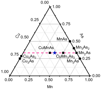
All known ternary phases in the Cu–Mn–As system have the transition metal () to As ratio of 2:1 and are either tetragonal or orthorhombic as shown in Fig. 1. The metallic nature of these compounds allows significant deviation from As stoichiometry, as evidenced by the binary compounds MnAs (a ferromagnet with a reentrant FeP-to-NiAs-type transition),Pytlik and Ziȩba (1985); Schwartz et al. (1971); Glazkov et al. (2003) Mn3As2 (which has at least three polymorphs),Dietrich et al. (1990); Möller and Jeitschko (1993); Hagedorn and Jeitschko (1994) the seemingly metastable compounds Mn4As3 and Mn5As4,Hagedorn and Jeitschko (1995); Möller and Jeitschko (1993) and Mn3As.Nowotny et al. (1951) Of these compounds, only MnAs and Mn2As have been investigated with neutron diffraction and transport measurements.Yuzuri and Yamada (1960); Austin et al. (1962) Further elaboration of compounds in this space is necessary to understand the potential for manipulating spins in these highly-correlated phases.
II Methods
Millimeter-sized crystals of hexagonal Cu0.82Mn1.18As were synthesized by mixing elemental powders Cu (99.9% metals basis), Mn (99.98% metals basis), and As (99.9999% metals basis) in 0.82:1.18:1 molar ratio. The powders were vacuum sealed in quartz tubes and heated at 1∘C/min to 600∘C for 6 hours then ramped at 1∘C/min to 975∘C for 1 hour. The tube was slow cooled at 1∘C/min to 900∘C and held for 1 hour before furnace-cooling down to room temperature. The resulting product was a solid ingot. The ingot was crushed into smaller pieces to conduct single crystal X-ray diffraction on a Bruker X8 Apex II diffractometer at 296 K and Å.
Variable-temperature powder X-ray diffraction was performed using a nitrogen blower at beamline 11-BM of the Advanced Photon Source in Argonne National Laboratory ( Å).Wang et al. (2008) Variable-temperature neutron powder diffraction was conducted at the WAND2 instrument at the High-Flux Isotope Reactor (HFIR) at Oak Ridge National Laboratory.Frontzek et al. (2018)
Magnetic structure determination was performed on a 2 mm crystal at the HB-3A four circle diffractometer at HFIR. A total of 344 reflections were collected at 4 K and used for structural refinement. Magnetic symmetry analysis was carried out using the tools available at the Bilbao Crystallographic ServerPerez-Mato et al. (2015) and refined using the FullProf suite.Rodríguez-Carvajal (1993)
Differential scanning calorimetry (DSC) measurements were performed on 5 mg of powder in Al pans under N2 atmosphere in a TA Instruments DSC 2500. A small fractured sample, weighing about 12 mg, was polished and aligned using Laue diffraction. This sample was mounted onto a quartz paddle sample holder for aligned magnetometry measurements in a Quantum Design MPMS3. Aligned resistivity measurements were carried out using the 4-point probe method in a Quantum Design PPMS DynaCool.
First-principles density functional theory (DFT) simulations were performed using the Vienna Ab-Initio Simulation Package (VASP) Kresse and Furthmüller (1996); Kresse and Joubert (1999). The electron-ion interaction is described using the projector-augmented wave (PAW) scheme.Blöchl (1994) Exchange and correlation are described using the generalized-gradient approximation (GGA) by Perdew, Burke, and Ernzerhof (PBE).Perdew et al. (1996) Single-particle Kohn-Sham states are expanded into a plane-wave basis with a cutoff energy of 600 eV. Monkhorst-Pack Monkhorst and Pack (1976) (MP) -point grids of and are used to integrate the Brillouin zone for cell relaxation and electronic band structure calculations, respectively. Non-collinear magnetism and spin-orbit coupling is taken into account in all calculations Steiner et al. (2016). Self-consistent total-energy convergence was achieved to within eV and atomic positions were relaxed until Hellman-Feynman forces were smaller than 5 meV/Å.
III Results and Discussion
III.1 Structure refinement
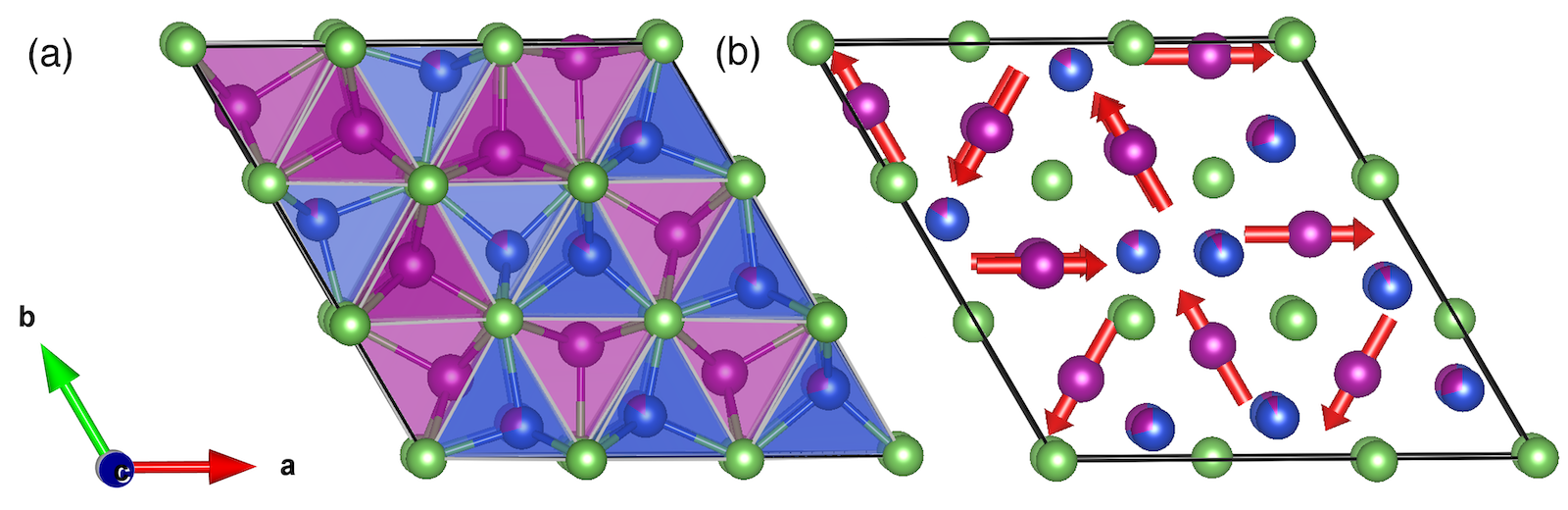
The refined structure of Cu0.82Mn1.18As is shown in Fig. 2(a), with structural parameters from single-crystal X-ray diffraction (XRD) given in Table 1 and 2. Cu0.82Mn1.18As has a short lattice parameter Å, indicating that the unit cell is flat and is the same width as the Cu and Mn coordination polyhedra. Fig. 2(a) shows the unit cell viewed down , with all the atoms occupying either = 0 or = 0.5. The compound forms in a new structure type with space group , and is comprised of three inequivalent square-pyramidal Mn and three inequivalent tetrahedral Cu, all coordinated by As. All metal sites have a multiplicity of 3 and have point symmetry. The atomic positions are well-described by the single-crystal XRD data, but the occupancies are less reliable due to the similar electron densities at each site. High-resolution synchrotron powder X-ray diffraction is shown in Fig. 3(a), to confirm that these samples can be made highly pure with excellent crystallinity.
The occupancies are better constrained by neutron scattering, where Mn and Cu have more contrast in their scattering lengths ( and 7.718 fm, respectively).Sears (1992) Neutron powder diffraction data from WAND2 were collected at 400 K, in the paramagnetic regime, with the refinement shown in Fig. 3(b). No evidence for site mixing or vacancies on the Mn or As sites was apparent. The best refinements were obtained by using the nominal Cu/Mn ratio and allowing Mn mixing on the Cu sites, with the final Cu occupancies of 0.709(2), 0.914(3), and 0.846(2) for Cu sites 1 – 3, respectively. The final structural refinement data presented in Table 2 is a single-crystal XRD refinement with the occupancies locked to values obtained by co-refinement to the 100 K synchrotron X-ray and 400 K neutron scattering data.

| Formula | Cu0.82Mn1.18As |
|---|---|
| Formula Weight | 191.88 g/mol |
| Crystal system | Hexagonal |
| Space group | |
| 11.1418(3) Å | |
| 3.8311(2) Å | |
| , | 411.87(3) Å3, 9 |
| 6.962 g/cm3 | |
| Absorption coefficient | 35.046 mm-1 |
| (000) | 777 |
| 0.714 | |
| Reflections collected | 6722 |
| Observed reflections | 953 |
| 0.0682 | |
| Number of parameters | 56 |
| Goodness-of-fit on | 1.445 |
| 0.0344, 0.0849 |
| Atom | Site | Occupancy | |||||||
|---|---|---|---|---|---|---|---|---|---|
| Cu1/Mn1 | 3j | 0.2489(9) | 0.0868(10) | 0 | 0.709/0.291(2) | 0.025(3) | 0.023(4) | 0.030(3) | 0.014(2) |
| Cu2/Mn2 | 3j | 0.5894(9) | 0.5043(5) | 0 | 0.914/0.086(3) | 0.012(3) | 0.011(2) | 0.015(2) | 0.005(3) |
| Cu3/Mn3 | 3k | 0.4206(9) | 0.5030(6) | 0.5 | 0.846/0.154(2) | 0.016(3) | 0.012(3) | 0.018(3) | 0.009(2) |
| Mn4 | 3j | 0.1966(10) | 0.4682(10) | 0 | 1 | 0.016(4) | 0.017(4) | 0.009(3) | 0.011(3) |
| Mn5 | 3k | 0.8051(10) | 0.9422(7) | 0.5 | 1 | 0.015(3) | 0.017(2) | 0.017(3) | 0.009(3) |
| Mn6 | 3k | 0.8056(10) | 0.5310(10) | 0.5 | 1 | 0.009(3) | 0.013(3) | 0.011(3) | 0.006(2) |
| As1 | 3j | 0.3310(6) | 0.3354(6) | 0 | 1 | 0.010(2) | 0.008(3) | 0.013(3) | 0.0044(19) |
| As2 | 3k | 0.6754(6) | 0.6721(6) | 0.5 | 1 | 0.010(2) | 0.013(3) | 0.0073(14) | 0.007(2) |
| As3 | 1a | 0 | 0 | 0 | 1 | 0.011(2) | 0.011(2) | 0.007(4) | 0.0054(12) |
| As4 | 1d | 1/3 | 2/3 | 0.5 | 1 | 0.007(2) | 0.007(2) | 0.012(4) | 0.0033(12) |
| As5 | 1e | 2/3 | 1/3 | 0 | 1 | 0.009(3) | 0.009(3) | 0.002(4) | 0.0045(14) |
III.2 Magnetic ordering
In light of the strong exchange coupling in transition-metal arsenides that leads to high Curie and Néel temperatures in MnAs and Mn2As, it is surprising that few hexagonal arsenides have been shown to order magnetically near room temperature. The most well-known structure type is the NiAs-type, of which MnAs is a member. NiAs itself is a Pauli paramagnet,Nozue et al. (1997) while hexagonal CrNiAs has a Curie temperature of 190 K.Stadnik et al. (2008)
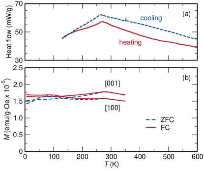
Powders of Cu0.82Mn1.18As were examined by DSC, with the heating and cooling traces shown in Fig. 4(a). There is a clear change in slope around 267 K, with a hysteresis of about 4 K. To determine the origin of this transition, aligned single crystals of Cu0.82Mn1.18As were examined via SQUID magnetometry, and the moment versus temperature is shown in Fig. 4(b). The maximum in the magnetometry data is around 275 K for zero-field-cooled (ZFC) and field-cooled (FC) data along the and axes for 10 kOe applied field. There are not sufficient data above to provide a satisfactory Curie-Weiss fit. The data along the axis display a typical decrease upon cooling past , while the data measured along the axis show a slight rise and plateau around 100 K. There were no features in measurements of magnetic moment versus field to indicate spin-flop transitions or any hysteresis. The small plateau could arise from decreasing itineracy and a leveling-off of the local moments on Mn sites, which would be consistent with the single-crystal neutron magnetic intensity remaining constant below 100 K. Fig. 4(b) shows that for temperatures beyond 150 K, the susceptibility along is larger than along . This trend is consistent with in-plane moments in triangular antiferromagnets such as CsMnBr3, CsVCl3, Mn3Sn etc.Brown et al. (1990); Kotyuzhanskii and Nikiforov (1991); Hirakawa et al. (1983); Duan et al. (2015) Below 150 K, the difference in the susceptibility along and is unclear. However, the in-plane Mn spin ordering was confirmed using neutron diffraction as shown below.
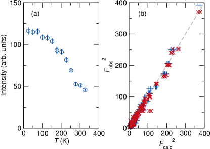
The magnetic ordering was probed first by variable-temperature neutron powder diffraction on the WAND2 instrument, which showed changes in peak intensities across this boundary, but no new peaks, indicating likely ordering. A full triple-axis data collection was performed on the HB-3A beamline at 4 K. The magnetic and nuclear structures were refined together in the magnetic space group. The intensity of the (020) peak can serve as an order parameter, and its temperature dependence is shown in Fig. 5(a). The (020) peak is an allowed nuclear reflection, so the intensity does not go to zero above . The three inequivalent Mn sites are constrained to have equal magnetic moments, which are refined to 3.02(8) /atom. No improvement in the fit was observed when the moments were allowed to freely vary. The observed and calculated structure factors are plotted in Fig. 5(b). The magnetic structure is shown in Fig. 2(b). No local Mn moment was stably refined on the Cu-majority sites, and Cu itself does not host local moments in arsenides Pauwels et al. (1973); Sampathkumaran et al. (2003); Sengupta et al. (2005). It is possible that some local Mn moments exist on the minority Cu sites, but they do not appear to be ordered. The 120∘ spin structure differs from Mn3Sn. In Mn3Sn, the spin triangles are connected by their corners. We also do not observe the “inverse triangle” orthorhombic configuration seen in Mn3Sn.Brown et al. (1990) There are three different types of 120∘ spin structures observed in our compound, although the spin directions in the plane could not be uniquely determined by unpolarized neutron diffraction. This compound could be written as containing Cu+ and Mn2+, but like other transition-metal arsenides the local moment is reduced due to metallicity.Katsuraki and Achiwa (1966); Pytlik and Ziȩba (1985)
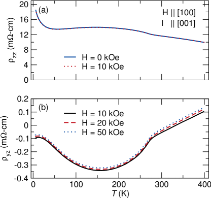
Four-point probe resistivity measurements along [001] show a mostly flat, weakly undulating trend versus temperature as shown in Fig. 6(a). The broad hump between 50 K to 250 K could be attributed to competing mobilities and carrier concentrations of multiple excited states in a heavily doped semiconductor (as in P-doped Si),Chapman et al. (1963) or variations in the dominant carrier scatterers in a disordered metal (which we discuss subsequently to be more likely, given the computed band structure). The resistivity values are roughly 125 times higher at 5 K in Cu0.82Mn1.18As than Fe2As and about 380 times higher than tetragonal CuMnAs, both of which are metallic Takeshita et al. (2017); Wadley et al. (2013). Application of a magnetic field of 10 kOe along [100] resulted in negligible change in resistivity values, shown in Fig. 6(a). A slightly larger effect can be seen in the Hall effect measurements in Fig. 6(b). The Hall data magnifies the hump around 150 K, and crosses from negative (majority -type) to positive (-type) upon heating past 330 K. The material is -type at low temperatures but as temperature is increased, more carriers are excited and the higher mobility of holes leads to compensation and switching to -type conduction 330 K. The lack of an anomaly in the total resistivity around the Hall crossover point indicates that the transport in Cu0.82Mn1.18As occurs via multiple bands, and is supported by the delicate (but not gapped) band structure around the Fermi energy that we discuss subsequently.
III.3 First-principles simulations
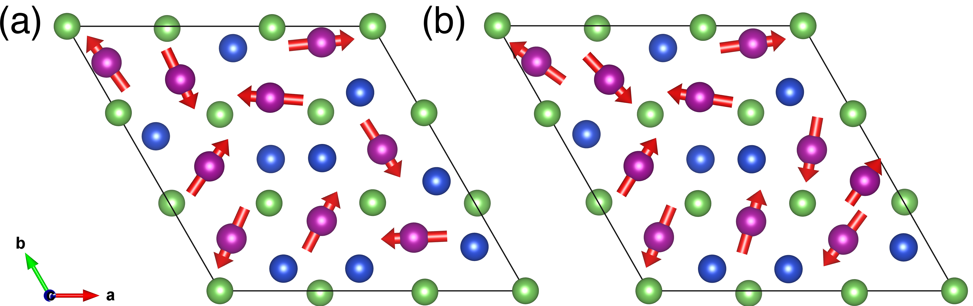
We performed first-principles density-functional theory (DFT) simulations to confirm the stability, cell geometry, and magnetic ordering of a fully-occupied hexagonal model compound CuMnAs and off-stoichiometric Cu0.89Mn1.11As, with a single Mn on a Cu1 site (1 of the 9 sites substituted per cell). We find that the relaxed atomic geometries of hexagonal CuMnAs and Cu0.89Mn1.11As agree with neutron scattering results within . The DFT data for the magnetic structures arrive at different lowest-energy orderings than the neutron refinement. The DFT-derived lowest-energy magnetic configurations of stoichiometric CuMnAs and substituted Cu0.89Mn1.11As are shown in Figs. 7(a) and (b), respectively. The stoichiometric result is antiferromagnetic, while the substituted site in Cu0.89Mn1.11As has a small uncompensated moment ( µB). The calculated neutron diffraction structure factors for the stoichiometric case are compared to the single-crystal neutron-refined values in Fig. 5(b). The two fits are similar, apart from the trio of peaks with , which significantly degrade the fit versus the neutron result. The neutron refinement outperforms the DFT fit with and versus and , respectively, where smaller numbers indicate a better fit. A small uncompensated moment observed in the Mn-substituted DFT model is an unavoidable artifact of the cell choice, which contains one “extra” Mn atom to reflect the off-stoichiometry of Cu0.82Mn1.18As.
Magnetic ground states in strongly-correlated -electron systems are often challenging to predict using DFT, so it is instructive to quantitatively evaluate the proximity of the neutron-refined result to the DFT energy minimum, and likewise the predicted neutron intensities of the DFT model. To better understand the energetics of this difference between theory and experiment, we compare total energies for three different situations: First, chemical and magnetic structures are constrained to the neutron scattering result ( in Table 3). Second, the ground-state magnetic structure is computed from DFT while the atomic geometries are constrained to the neutron scattering data ( in Table 3). Finally, these total energies are compared to the fully relaxed DFT result ( in Table 3). These small energy changes, 15.30 and 14.93 meV/atom for CuMnAs and Cu0.89Mn1.11As, respectively are typical of energy differences between various magnetic structures for similar systems.Lutz (2013)
| System | |||||
|---|---|---|---|---|---|
| CuMnAs | 9.92 | 15.30 | 11.050 | 11.050 | 3.802 |
| Cu0.89Mn1.11As | 6.45 | 14.93 | 11.053 | 11.043 | 3.776 |
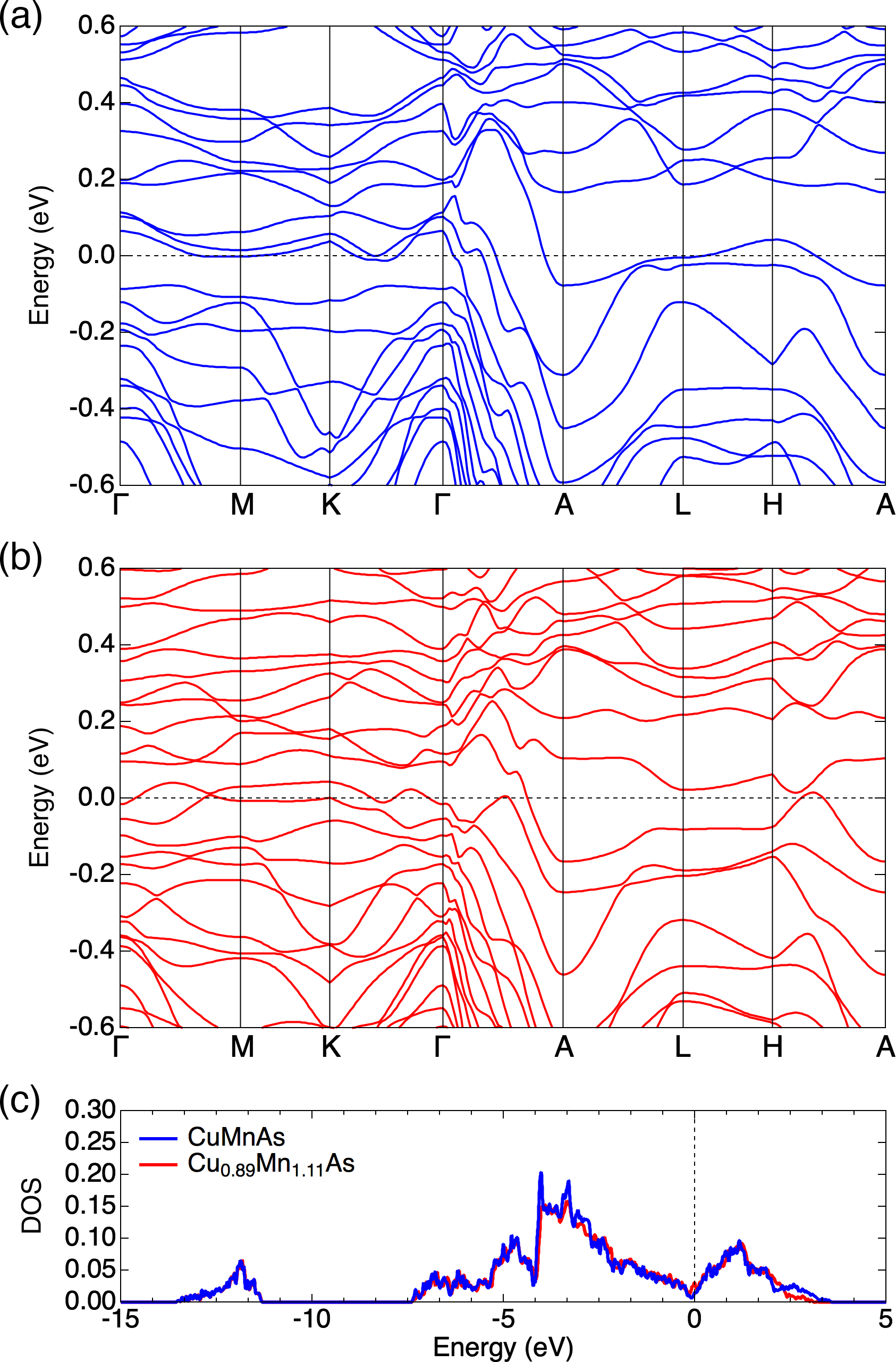
The electronic band structure and density of states of stoichiometric hexagonal CuMnAs and Cu0.89Mn1.11As in Fig. 8 show that both hexagonal models are metallic. Both electronic structures exhibit very small densities of states near the Fermi energy, similar to that described by DFT for tetragonal CuMnAs Máca et al. (2017). Tetragonal CuMnAs shows obvious metallic resistivity ().Wadley et al. (2013) The CuMnAs compounds are clearly on the cusp of semiconducting/metallic behavior, and share similarities to Fe2As, which has a much greater density of states at the Fermi level and does show , but the reported values of resistivity values are much higher than that of tetragonal CuMnAs.Yang et al. (2019); Takeshita et al. (2017)
Our DFT calculations suggest that Cu0.89Mn1.11As is metallic. However, transport measurements indicate that the resistivity is high, and for most . The negative slope that is observed at low and high temperatures is not exponential as is expected in highly-doped semiconductors.Chapman et al. (1963) The seeming discrepancy between resistivity and the computed band structure can be resolved by considering the high amount of substitutional disorder in these compounds. Metals often exhibit behavior when a large amount of configurational disorder is present,Elk et al. (1979) and the negative temperature dependence is in fact correlated with high absolute values of resistivity.Mooij (1973) In our material, carriers must scatter due to pervasive disorder due to Mn site mixing, while magnon scattering may also contribute strongly, but the overall resistivity is hardly affected upon cooling past .
IV Conclusions
We report the crystal structure of a non-centrosymmetric phase in the Cu–Mn–As system, with a new structure type. This compound can be made phase-pure in single crystal form. Triangular antiferromagnetic ordering appears upon cooling below 270 K and is markedly distinct from the orthorhombic and tetragonal CuMnAs phases, both of which are stabilized by different Cu/Mn content and are centrosymmetric in their paramagnetic states. DFT calculations confirm the stability of the magnetic structure refined by single-crystal neutron diffraction. The triangular AF ordering is in-plane and does not break degeneracy of the and axes. Like other copper manganese arsenides, hexagonal Cu0.82Mn1.18As is on the cusp of semiconducting/metallic behavior and further investigation of the carrier scattering mechanisms in this class of materials is warranted.
V Acknowledgments
This work is supported by the National Science Foundation (NSF) under Grant No. DMR-1720633. Characterization was carried out in part in the Materials Research Laboratory Central Research Facilities, University of Illinois. Use of the Advanced Photon Source at Argonne National Laboratory was supported by the U.S. Department of Energy, Office of Science, Office of Basic Energy Sciences, under Contract No. DE-AC02-06CH11357. Neutron scattering was performed at the High Flux Isotope Reactor, a Department of Energy Office of Science User Facility operated by the Oak Ridge National Laboratory. This work made use of the Illinois Campus Cluster, a computing resource that is operated by the Illinois Campus Cluster Program (ICCP) in conjunction with the National Center for Supercomputing Applications (NCSA) and which is supported by funds from the University of Illinois at Urbana-Champaign. We thank Junseok Oh for assistance in making contacts for resistivity measurements.
References
- Wadley et al. (2016) P. Wadley, B. Howells, J. Zelezny, C. Andrews, V. Hills, R. P. Campion, V. Novak, K. Olejnik, F. Maccherozzi, S. S. Dhesi, S. Y. Martin, T. Wagner, J. Wunderlich, F. Freimuth, Y. Mokrousov, J. Kunes, J. S. Chauhan, M. J. Grzybowski, A. W. Rushforth, K. W. Edmonds, B. L. Gallagher, and T. Jungwirth, Science 351, 6273 (2016).
- Grzybowski et al. (2017) M. J. Grzybowski, P. Wadley, K. W. Edmonds, R. Beardsley, V. Hills, R. P. Campion, B. L. Gallagher, J. S. Chauhan, V. Novak, T. Jungwirth, F. Maccherozzi, and S. S. Dhesi, Phys. Rev. Lett. 118, 057701 (2017).
- Wadley et al. (2018) P. Wadley, S. Reimers, M. J. Grzybowski, C. Andrews, M. Wang, J. S. Chauhan, B. L. Gallagher, R. P. Campion, K. W. Edmonds, S. S. Dhesi, F. Maccherozzi, V. Novak, J. Wunderlich, and T. Jungwirth, Nat. Nanotechnol. 13, 362 (2018).
- Matalla-Wagner et al. (2019) T. Matalla-Wagner, M.-F. Rath, D. Graulich, J.-M. Schmalhorst, G. Reiss, and M. Meinert, arxiv (2019), arXiv:1903.12387 .
- Wadley et al. (2015) P. Wadley, V. Hills, M. R. Shahedkhah, K. W. Edmonds, R. P. Campion, V. Novák, B. Ouladdiaf, D. Khalyavin, S. Langridge, V. Saidl, P. Nemec, A. W. Rushforth, B. L. Gallagher, S. S. Dhesi, F. MacCherozzi, J. Železný, and T. Jungwirth, Sci. Rep. 5, 17079 (2015).
- Hills et al. (2015) V. Hills, P. Wadley, R. P. Campion, V. Novak, R. Beardsley, K. W. Edmonds, B. L. Gallagher, B. Ouladdiaf, and T. Jungwirth, J. Appl. Phys. 117 (2015).
- Uhlirova et al. (2015) K. Uhlirova, R. Tarasenko, F. J. Martinez-Casado, B. Vondrackova, and Z. Matej, J. Alloy Compd. 650, 224 (2015).
- Uhlirova et al. (2019) K. Uhlirova, E. Duverger-Nedellec, R. H. Colman, J. Volny, B. Vondrackova, and K. Carva, J. Alloy Compd. 771, 680 (2019).
- Nateprov et al. (2011) A. N. Nateprov, V. C. Kravtsov, V. Fritsch, and H. von Löhneysen, Surf. Eng. Appl. Elect. 47, 540 (2011).
- Máca et al. (2012) F. Máca, J. Mašek, O. Stelmakhovych, X. Martí, H. Reichlová, K. Uhlířová, P. Beran, P. Wadley, V. Novák, and T. Jungwirth, J. Magn. Magn. Mater. 324, 1606 (2012).
- Tang et al. (2016) P. Tang, Q. Zhou, G. Xu, and S. C. Zhang, Nat. Phys. 12, 1100 (2016).
- Kim et al. (2018) Y. Kim, K. Kang, A. Schleife, and M. J. Gilbert, Phys. Rev. B 97, 134415 (2018).
- Emmanouilidou et al. (2017) E. Emmanouilidou, H. Cao, P. Tang, X. Gui, C. Hu, B. Shen, J. Wu, S.-C. Zhang, W. Xie, and N. Ni, Phys. Rev. B 96, 224405 (2017).
- Pytlik and Ziȩba (1985) L. Pytlik and A. Ziȩba, J. Magn. Magn. Mater. 51, 199 (1985).
- Schwartz et al. (1971) L. H. Schwartz, E. L. Hall, and G. P. Felcher, J. Appl. Phys. 42, 1621 (1971).
- Glazkov et al. (2003) V. P. Glazkov, D. P. Kozlenko, K. M. Podurets, B. N. Savenko, and V. A. Somenkov, Crystallogr. Rep. 48, 59 (2003).
- Dietrich et al. (1990) L. H. Dietrich, W. Jeitschko, and M. H. Möller, Z. Kristallogr. Cryst. Mater. 190, 259 (1990).
- Möller and Jeitschko (1993) M. H. Möller and W. Jeitschko, Z. Kristallogr. Cryst. Mater. 204, 1 (1993).
- Hagedorn and Jeitschko (1994) M. F. Hagedorn and W. Jeitschko, J. Solid State Chem. 113, 257 (1994).
- Hagedorn and Jeitschko (1995) M. F. Hagedorn and W. Jeitschko, J. Solid State Chem. 119, 344 (1995).
- Nowotny et al. (1951) H. Nowotny, R. Funk, and J. Pesl, Monatsh. Chem. 82, 513 (1951).
- Yuzuri and Yamada (1960) M. Yuzuri and M. Yamada, J. Phys. Soc. Jpn. 15, 1845 (1960).
- Austin et al. (1962) A. E. Austin, E. Adelson, and W. H. Cloud, J. Appl. Phys. 33, 1356 (1962).
- Wang et al. (2008) J. Wang, B. H. Toby, P. L. Lee, L. Ribaud, S. M. Antao, C. Kurtz, M. Ramanathan, R. B. Von Dreele, and M. A. Beno, Rev. Sci. Instrum. 79, 085105 (2008).
- Frontzek et al. (2018) M. D. Frontzek, R. Whitfield, K. M. Andrews, A. B. Jones, M. Bobrek, K. Vodopivec, B. C. Chakoumakos, and J. A. Fernandez-Baca, Rev. Sci. Instrum. 89, 0928011 (2018).
- Perez-Mato et al. (2015) J. Perez-Mato, S. Gallego, E. Tasci, L. Elcoro, G. de la Flor, and M. Aroyo, Ann. Rev. Mater. Res. 45, 217 (2015).
- Rodríguez-Carvajal (1993) J. Rodríguez-Carvajal, Physica B 192, 55 (1993).
- Kresse and Furthmüller (1996) G. Kresse and J. Furthmüller, Phys. Rev. B 54, 11169 (1996).
- Kresse and Joubert (1999) G. Kresse and D. Joubert, Phys. Rev. B 59, 1758 (1999).
- Blöchl (1994) P. E. Blöchl, Phys. Rev. B 50, 17953 (1994).
- Perdew et al. (1996) J. P. Perdew, K. Burke, and M. Ernzerhof, Phys. Rev. Lett. 77, 3865 (1996).
- Monkhorst and Pack (1976) H. J. Monkhorst and J. D. Pack, Phys. Rev. B 13, 5188 (1976).
- Steiner et al. (2016) S. Steiner, S. Khmelevskyi, M. Marsmann, and G. Kresse, Phys. Rev. B 93, 224425 (2016).
- Sears (1992) V. F. Sears, Neutron News 3, 26 (1992).
- Nozue et al. (1997) T. Nozue, H. Kobayashi, M. Sato, A. Uesawa, T. Suzuki, and T. Kamimura, Physica B 237-238, 174 (1997).
- Stadnik et al. (2008) Z. M. Stadnik, P. Wang, N. Jansen, D. Walcher, P. Gütlich, and T. Kanomata, J. Phys. Cond. Mat. 20, 325230 (2008).
- Brown et al. (1990) P. J. Brown, V. Nunez, F. Tasset, J. B. Forsyth, and P. Radhakrishna, J. Phys.: Condens. Matter 2, 9409 (1990).
- Kotyuzhanskii and Nikiforov (1991) B. Y. Kotyuzhanskii and D. V. Nikiforov, J. Phys.: Condens. matter 3 (1991).
- Hirakawa et al. (1983) K. Hirakawa, H. Ikeda, H. Kadowaki, and K. Ubukoshi, J. Phys. Soc. Jpn. 52, 2882 (1983).
- Duan et al. (2015) T. F. Duan, W. J. Ren, W. L. Liu, S. J. Li, W. Liu, and Z. D. Zhang, Appl. Phys. Lett. 107 (2015).
- Pauwels et al. (1973) L. J. Pauwels, G. Maervoet, and R. Vervaeke, Z. Anorg. Allg. Chem. 397, 307 (1973).
- Sampathkumaran et al. (2003) E. V. Sampathkumaran, K. Sengupta, S. Rayaprol, K. K. Iyer, T. Doert, and J. P. F. Jemetio, Phys. Rev. Lett. 91, 036603 (2003).
- Sengupta et al. (2005) K. Sengupta, P. L. Paulose, E. V. Sampathkumaran, T. Doert, and J. P. F. Jemetio, Phys. Rev. B 72, 184424 (2005).
- Katsuraki and Achiwa (1966) H. Katsuraki and N. Achiwa, J. Phys. Soc. Japan 21, 2238 (1966).
- Chapman et al. (1963) P. W. Chapman, O. N. Tufte, J. D. Zook, and D. Long, J. Appl. Phys. 34, 3291 (1963).
- Takeshita et al. (2017) N. Takeshita, I. Akira, S. Ishida, H. Eisaki, and Y. Yoshida, J. Phys.: Conf. Ser. 950, 042024 (2017).
- Wadley et al. (2013) P. Wadley, V. Novák, R. P. Campion, C. Rinaldi, X. Martí, H. Reichlová, J. Zelezný, J. Gazquez, M. A. Roldan, M. Varela, D. Khalyavin, S. Langridge, D. Kriegner, F. Máca, J. Masek, R. Bertacco, V. Holy, A. W. Rushforth, K. W. Edmonds, B. L. Gallagher, C. T. Foxon, J. Wunderlich, and T. Jungwirth, Nat. Commun. 4, 2322 (2013).
- Lutz (2013) L. C. Lutz, Electronic structure, magnetic structure, and metal-atom site preferences in CrMnAs, Graduate theses and dissertations, Iowa State University (2013).
- Máca et al. (2017) F. Máca, J. Kudrnovsk’y, V. Drchal, K. Carva, P. Baláž, and I. Turek, Phys. Rev. B 96, 094406 (2017).
- Yang et al. (2019) K. Yang, K. Kang, Z. Diao, A. Ramanathan, M. H. Karigerasi, D. P. Shoemaker, A. Schleife, and D. G. Cahill, arXiv , 1 (2019), arXiv:1903.07810 .
- Elk et al. (1979) K. Elk, J. Richter, and V. Christoph, J. Phys. F 9, 307 (1979).
- Mooij (1973) J. H. Mooij, Phys. Status Solidi A 17, 521 (1973).