Estimating sex and age for forensic applications using machine learning based on facial measurements from frontal cephalometric landmarks
Abstract
Facial analysis permits many investigations some of the most important of which are craniofacial identification, facial recognition, and age and sex estimation. In forensics, photo-anthropometry describes the study of facial growth and allows the identification of patterns in facial skull development by using a group of cephalometric landmarks to estimate anthropological information. Previous works presented, as indirect applications, the use of photo-anthropometric measurements to estimate anthropological information such as age and sex. In several areas, automation of manual procedures has achieved advantages over and similar measurement confidence as a forensic expert. This manuscript presents an approach using photo-anthropometric indexes, generated from frontal faces cephalometric landmarks, to create an artificial neural network classifier that allows the estimation of anthropological information, in this specific case age and sex. The work is focused on four tasks: i) sex estimation over ages from 5 to 22 years old, evaluating the interference of age on sex estimation; ii) age estimation from photo-anthropometric indexes for four age intervals (1 year, 2 years, 4 years and 5 years); iii) age group estimation for thresholds of over 14 and over 18 years old; and; iv) the provision of a new data set, available for academic purposes only, with a large and complete set of facial photo-anthropometric points marked and checked by forensic experts, measured from over 18,000 faces of individuals from Brazil over the last 4 years. The proposed classifier obtained significant results, using this new data set, for the sex estimation of individuals over 14 years old, achieving accuracy values greater than 0.85 by the F1 measure. For age estimation, the accuracy results are 0.72 for measure with an age interval of 5 years. For the age group estimation, the measures of accuracy are greater than 0.93 and 0.83 for thresholds of 14 and 18 years, respectively.
Index Terms:
Forensics, Artificial Neural Network, Facial Photo-anthropometry, Computer vision, age and sex recognition, Anthropology.I Introduction
Anthropological knowledge can be used to support forensic investigations of the deceased and the living [1]. In the first case, postmortem (PM) profiles of victims are reconstructed to narrow the number of comparisons between missing persons and unknown bodies, as described in [2]. The profiling process is carried out by retrieving information regarding sex, age, stature and ancestry from the deceased, especially from skeletal remains, and comparing them with antemortem (AM) data from the alleged victim [3]. In order to increase reliability, the collected information is combined with AM and PM evidence obtained by primary means of human identification, namely fingerprint, dental and DNA analyses [4].
On the other hand, forensic anthropology applied to the living usually relies on morphological and biometric information of victims and suspects of crimes registered in footage of closed-circuit television and photographs [5, 6]. The identification of children that suffered sexual exploitation, as well as their perpetrators, figures among the procedures requested by Law in the routines of medico-legal institutes [7]. Over the last decade, requests for anthropological examination of the living became more common following an increasing trend in cybernetic crimes [8]. This new outlook justified the need for developing advanced tools to support forensic casework [9].
The photo-anthropometric analysis of the human face emerges in this context as an alternative tool for searching, collecting, and quantifying morphological features and using them for forensic purposes [10]. Working at the interface of forensic anthropology and computer science, this non-invasive and low-cost approach is founded on the registration of landmarks on photographs and the calculation of ratios between facial distances [7, 9, 10]. The morphometric information retrieved from the human face can be used within a comparative basis, between reference and target persons, or in a reconstructive basis, where sexual dimorphism and age estimation of the living are performed [11].
This study was designed with four aims for the use of photo-anthropometric data of the human face: to propose an automatic solution based on an artificial neural network to estimate anthropological information using photo-anthropometric indices (I); to test the diagnostic accuracy of the solution with cut-off points between male and females and threshold limits for the ages of 14 - related to sexual consent [12, 13], and 18 - related to legal majority [14, 15] (II); to analyze the correlation between sex and age using photo-anthropometric indices of the human face (III) and; provision, for academic purposes only, of a complete data set of facial photo-anthropometric points marked and checked by forensic experts, measured on over 18.000 faces of individuals from Brazil over the last 4 years (IV).
The manuscript is organized as follows: Section II presents the proposed method and all the processing details for classification using a model based on artificial intelligence techniques. It also includes a description of the database and the inputs used in this manuscript. Section III presents the experimental results. Finally, Section IV presents the discussion, main conclusions and future works.
II Materials and Methods
The main goal of this manuscript is to evaluate the use of photo-anthropometric data from human faces to create an automatic classifier based on an artificial neural network to estimate age and sex. In this Section we will describe the approach used to create an automatic solution using an artificial intelligence model and the details of the inputs and the proposed tests.
II-A Photos Database and Cephalometric Landmarks sets
The principal proposed and used database is composed of photo-anthropometric index data from 18,000 frontal face photos of 18,000 different people from Brazil. All photos were acquired in accordance with ICAO 9303 normative [16] used for Machine Readable Travel Documents. All photos have been captured over a white background, stored at a resolution of 480x640 pixels, 24bits of color, with no glasses and with a natural expression. The photos are divided into male (9,000) and female (9,000) and 18 age groups (from 5 up to 22 years old), totaling 500 photos for each individual group (sex and age).
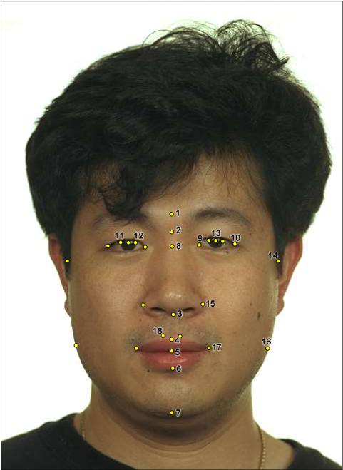
The cephalometric landmarks adopted in this manuscript, all of which were used to create the photo-anthropometric indexes, were described in [9] and [7]. In this methodology, one expert manually located 28 cephalometric landmarks in each of a 1000 pre-training images using a SAFF-2D software, applying an identification methodology proposed in [10, 17, 18]. All 18,000 faces were processed and all facial landmarks were marked and checked following the methodology developed in [19]. The following is a complete list of cephalometric landmarks which were identified, according to Caple and Stephan standard nomenclature [20]: Entocanthion (en’), Exocanthion (ex’), Iridion laterale (il), Iridion mediale (im), Pupil (pu’), Zygion (zy’), Alare (al’), Gonion (go’) and Cheilion (ch’), Crista philtri (cph’) bilateral landmarks. The remaining can be found on face’s midline as: Glabella (g’), Nasion (n’), Subnasale (sn’), Labiale superius (ls’), Stomion (sto’), Labiale inferius (li’), Gnathion (gn’), Midnasal (m’) [21]. Figure 1 shows all the landmarks used and their locations on the face.
II-B Photo-anthropometric indexes
Using the cephalometric landmarks detailed in Subsection II-A, the authors of [9] and [7] present details about 10 and 40 facial photo-anthropometric indexes (PAIs) respectively. They are facial measures which permit the explanation of people’s anthropometric information, specifically age and sex, by only using facial growth information.
The main feature of the PAIs is the use of the mean iris diameter as a proportional factor defined as the iris ratio to solve scale and calibration issues (e.g. different metrics relations to the pixel size and to the intrinsic parameters of the capture device which was used). The iris ratio is essentially the Euclidean distance defined by a function from Iridion laterale (il) and Iridion mediale (im) of both eyes. The iris ratio is detailed in Equation 1. When a facial measurement generated by the Euclidean distance between two cephalometric landmarks is divided by the iris ratio the result is a stable growing factor which solves the image scale problem and removes effects of varying landmark positions over the face.
| (1) |
Using a specialized software to automatically extract the cephalometric landmarks, we applied the methodology (iris ratio) proposed in [9], combining all of the 28 cephalometric landmarks creating 208 PAIs. The result is stored in a comma-separated values file (CSV) composed of the photo-anthropometry data, including 208 PAIs per image, labeled by sex and age for the 18.000 images used to build the data set. More details about the generated data set will be described in Section II-D. The complete description of all 208 PAIs can be found in the supplementary material in Section B.
II-C Experimental set-up and evaluation metrics
All the proposed tests were executed on an Intel AI DevCloud [23], a cloud framework for applications in artificial intelligence powered by Intel, running the Keras API version 2.2.0 [24] and Tensorflow version 1.8 [25]. During the training process we adopted an artificial neural network (ANN) model based on a multilayer perceptron (MLP) [26], using one dense (fully connected) layer with 128 neurons with the Adamax optimizer from Adam [27]. Table I presents the detailed MLP structure used to execute the tests.
| # Input layer | 209 (208 PAIs + sex attribute) |
|---|---|
| # Hidden layer | 1 |
| # Neuron in hidden layer | 128 |
| # Epochs | 500 |
| Activation function in hidden layer | Sigmoid |
| Activation function in output layer | Softmax |
| Learning rate | 0.01 |
| Momentum | 0.9 |
| Optimizer | Adamax |
In a classification process, all results require a test accuracy analysis and a traditional measure is the F1 score (also known as F-score or F-measure) [28]. This accuracy metric is composed of four parameters: True Positives (), True Negatives (), False Positives () and False Negatives (). At the end of the estimation process, we evaluated the estimate as a true positive () when the classifier agreed with the anthropological data (age/sex) for the validation data and as (false positive) when the estimate (age/sex) disagreed. We have used the F1 score method shown in Equation 2,
| (2) |
where the variables and are defined as and , respectively. The F1 score metric varies between 1 (best) and 0 (worst). To evaluate each test in detail, we adopted the confusion matrix method [29]. The confusion matrix allows the comparison of each “predicted label” with the “true label” resulting in a table which presents the classifier’s behavior, providing a more complete visualization than the F1 score.
As described above, we propose the use of 208 PAIs per photo and provide them as input to an automatic classifier to estimate anthropological information. In order to evaluate the expected performance of the classifier model over the proposed data set, a k-fold cross-validation was used in the testing process [30, 31]. In our case, a ten-fold cross-validation procedure was used, randomly separating our data set five times with of photos for training the classifiers and of photos for testing.
To evaluate the PAI methodology for anthropological estimation, we defined 3 groups of tests. The first one, Group A relates to the estimation of sex, the second one, Group B, to the estimation of age and Group C to the estimation of the age group. These tests are described as follows:
-
•
Group A - Sex estimation: The group is composed of two test sets where one classifier was trained with age as an input and the other classifier avoiding111When the term “avoiding” is used throughout the manuscript, it means information from a specific class was not used in the proposed tests. it, working similarly to an adversarial neural network [32].
-
•
Group B - Age estimation: This group is composed of 4 tests to evaluate age estimation using only the PAI data. For all these tests, we focus on identifying if the sex data can provide improvements over (or interfere with) the age estimation using only the facial measurements.
-
•
Group C - Group age estimation: In this group two classifiers are proposed in order to evaluate if the facial measurements are able to identify if an individual belongs to a specific age group. The classifiers analyze the thresholds of 14 and 18 years old. The first one identifies if the person is 14 years old or more and the second one if the person is 18 years old or older.
As stated above, in Group A the estimation of sex is analyzed. Table II presents the structure of the tests for Group A. In Test 1, we trained the classifier separately for each age group, totaling 17 individual tests from 5 up to 22 years old. This test was developed to try to evaluate the classification process applied on each age range using only the PAI data. In Test 2 we used all the data observations (avoiding the age information) to evaluate the sex classification over all age groups.
| Test | Target | Age as input | Qty. Test |
|---|---|---|---|
| 1 | Sex | Yes | 17 |
| 2 | Sex | No | 1 |
Meanwhile in Group B the age estimation process is analyzed. Table III presents the test structure of the Group B tests. We defined 4 tests to evaluate the classification process for age estimation using the PAI data. For each test we analyzed the impact of sex information on the estimation process.
| Test | Target | Sex as input |
|---|---|---|
| 1 | 6,7,8,9,10,11,12,13,14,15,16,17,18,19,20,21,22 | No/Female/Male |
| 2 | 6,8,10,12,14,16,18 ,20,22 | No/Female/Male |
| 3 | 6,10,14,18,22 | No/Female/Male |
| 4 | 5,10,15,20 | No/Female/Male |
For each proposed test, we trained three different classifiers. Two by splitting the PAI data by sex (female and male) and the other classifier using all the data observations in the same classification process. The 4 tests in Group B are described as follow:
-
•
Test 1: We selected the PAI data from 6 to 22 years old at age intervals of 1 year. In this test we are focused on evaluating the classifier accuracy for age estimation with a total of 17 age classes as output.
-
•
Test 2: We selected the PAI data from 6 to 22 years old at age intervals of 2 years. In this test, as in Test 1, we evaluate the classifier accuracy for age estimation with 9 age classes as output: 6, 8, 10, 12, 14, 16, 18, 20 and 22.
-
•
Test 3: We selected the PAI data from 6 to 22 years old at age intervals of 4 years. As above, we are evaluating the classifier accuracy for age estimation with a total of 5 age class outputs: 6, 10, 18 and 22.
-
•
Test 4: We selected the PAI data from 5 to 20 years old at age intervals of 5 years. As in all the previous tests of this group, we are interested in evaluating the classifier accuracy for age estimation with a total of 4 output classes: 5, 10, 15 and 20.
Finally, for Group C, Table IV presents the test structure for age group classification, with thresholds of 14 and 18 years. In these tests we evaluated if the PAIs can be used to estimate if the person is older/under 14 years old or older/under 18 years old. We adopted the same test procedures as presented in Group B. In this case we evaluated if the sex data affects the age group estimation process. For both tests we trained classifiers for females, for males and for all observations without the sex information as input.
| Test | Target | Sex as input |
|---|---|---|
| 1 | Older/under 14 years | No/Female/Male |
| 2 | Older/under 18 years | No/Female/Male |
II-D Descriptive Statistical Analyses of the Data Set
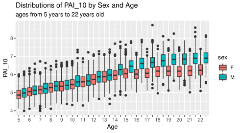
In this section we undertake a descriptive statistical exploration of the new data set which serves as input to the proposed machine learning model222The data set and the developed machine learning model will be available to download soon, after the blind review process is completed, in attendance of the journal’s submission criteria.. As described above, the full data set contains 500 observations on 208 variables for each category of Age (18 levels) and Sex (2 levels), yielding observations in total. Given the large number of variables and categories, it is not a viable option to describe all the variable distributions in all the categories. Therefore, we performed a visual and detailed analysis of boxplots of the distributions of all the variables in all the 36 categories. An example for the variable PAI-10 (wing of the nose - chin) can be found in Figure 2. Males present larger values than females and the differences increase with age. Similar figures for all 208 variables can be found in the supplementary material333All boxplots are available in the specific section of the Supplementary Material files (File PAIs_sex_age_boxplots_supplemental.pdf)..
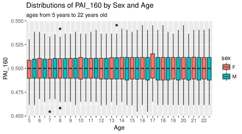
We performed Shapiro-Wilk Normality Tests [33] in each age-sex group for each variable. The null hypothesis for this test is that the data are normally distributed. values lower than were considered indications of significant deviations from normality. Three variables were non-normal in all age-sex groups: PAI-154 (diameter of the iris), PAI-160 (lateral iris - pupil), and PAI-171 (medial iris - pupil, same side). For the other variables, approximately of the tests rejected normality (about two age-sex groups per variable). However, given the large number of observations in each category and the presence of many outliers444See Figure 2, for example, where outliers can be identified as the points well above or well below the boxes in the boxplot., both of which increase the probability of rejecting normality, this suggests that much of the data is normally distributed and that analysis techniques that depend on normality can be used, albeit with caution.
Two-way analyses of variance (ANOVA) [34] were performed on each PAI to assess statistically significant changes in facial parameters in response to the factors sex, age, and the interaction between sex and age. values lower than were considered significant. For each of the 208 PAIs both sex and age were significant factors. The interaction between sex and age was not significant for only 4 of the 208 PAIs, specifically PAI-50 (labial commissure - zygion, same side), PAI-154 (diameter of the iris), PAI-160 (lateral iris - pupil), and PAI-171 (medial iris - pupil, same side). Three of these four variables were identified as non-normal in all categories by the Shapiro-Wilk tests presented above, only PAI-50 was not. As an example, the distributions of PAI-160 are presented in Figure 3. It is interesting to observe that the inclusion of these variables in the proposed machine learning model does not introduce any difficulties, as would occur in a standard regression model where large correlations and/or lack of variation would cause instability in the model estimates. The model simply will not use variables which don’t provide additional predictive power.
III Results
This section presents all the results obtained using the proposed artificial neural network architecture detailed in Subsection II-C, including all the F1 scores and confusion matrices for each proposed test.
III-A F1 results
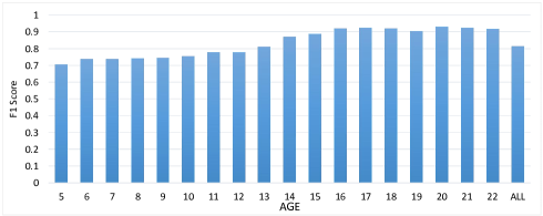
For Group A, the evaluated tests describe how the PAI indexes can classify the sex information. Figure 4 presents the F1 score for sex estimation for each age. The last column (all) was obtained using all the ages in the classification process in order to evaluate the sex classification baseline test or “global” test. We can compare the mean of the results for each age to the “global” test, obtaining similar F1 scores: and respectively.
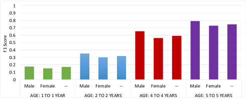
The tests from Group B use the PAI indexes for age classification. Figure 5 presents the F1 scores for age estimation divided in four groups: age intervals of 1 year (green), age intervals of 2 years (blue), age intervals of 4 years (red) and age intervals of 5 years (purple). Each group test was divided into three subtests: the first one is the age estimation on male individuals. The second one just female individuals and the last one using both and avoiding the sex information.
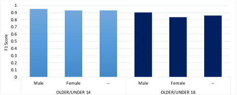
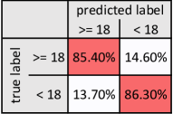
The Group C tests are focused on evaluating if the PAI indexes can classify the age group information correctly. Figure 6 presents the F1 scores for age group estimation divided into two tests: older/under 14 years old and older/under 18 years old. Each group test was divided into three subtests, as were the Group B tests: male individuals; female individuals; and all individuals avoiding the sex information.
III-B Confusion Matrices
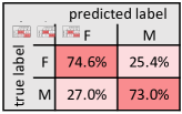
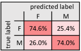
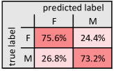
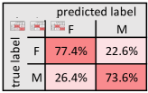
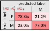
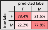
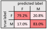
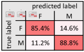
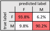
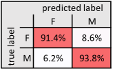
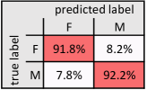
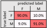
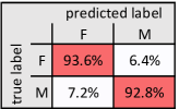
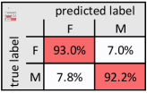
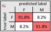
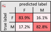
For each group test, we used confusion matrices to provide a detailed report, including all achieved results for age and sex classification. The matrices demonstrate the classifiers’ accuracy by comparing the “predicted labels” to the “true labels”.
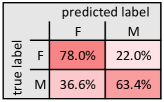
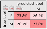
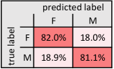
Figure 7 presents the confusion matrices for the Group A tests. The figures from Figure 7 LABEL:sub@fig:mf_age_1 to Figure 7 LABEL:sub@fig:mf_age_22 present the confusion matrices for each age group separately, from 5 to 22 years old respectively. Figure 7 LABEL:sub@fig:mf_age_avg presents the average results of all age groups, meanwhile the Figure 7 LABEL:sub@fig:mf_age_all presents the results using all data avoiding the age information.

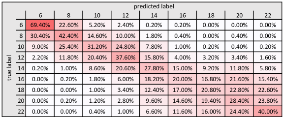
Figures 8, 9 and 10 present the confusion matrices for tests in Group B using 1 year for age intervals. Figure 8 presents the results for age estimation avoiding the sex information. Figure 9 presents the results for age estimation for females. Figure 10 presents the results for age estimation for males.
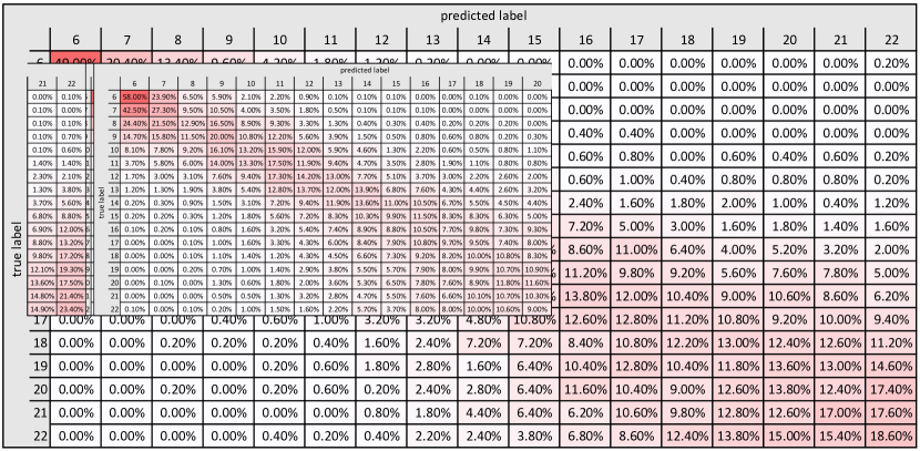
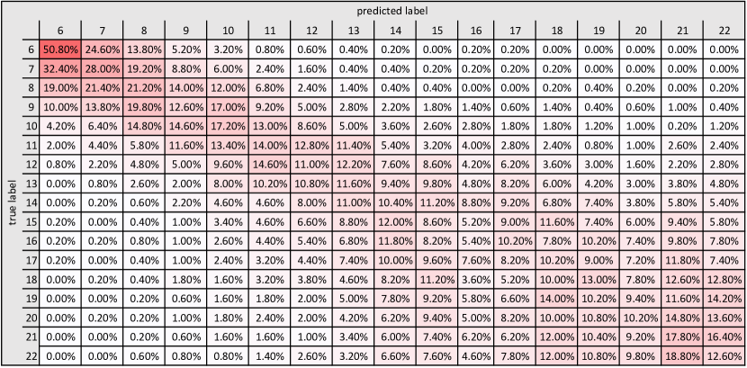
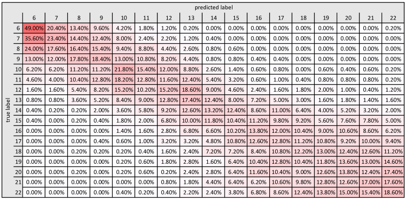
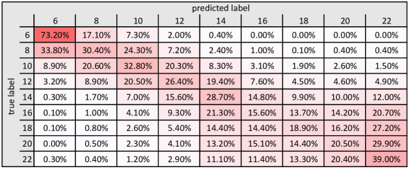
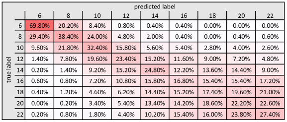
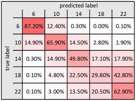
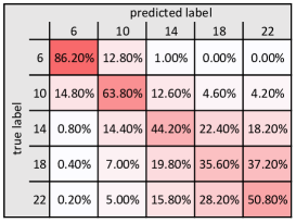
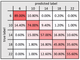
Figures 13, 14 and 15 present the confusion matrices with the detailed results for Group B: age intervals of 2 years. Figure 13 presents the detailed results for age estimation avoiding the sex information. Figure 14 and Figure 15 present the detailed results for age estimation for female and male individuals, respectively.
Figure 16 presents the confusion matrices with the detailed results for Group B: age intervals of 4 years. Figure 16 LABEL:sub@fig:age_4_4 presents the detailed results for age estimation avoiding the sex information. Figure 16 LABEL:sub@fig:age_4_4_F presents the detailed results for age estimation on female individuals. Figure 16 LABEL:sub@fig:age_4_4_M presents the detailed results for age estimation on male individuals.
Figure 17 presents the confusion matrices with the detailed results for Group B: age intervals of 5 years. Figure 17 LABEL:sub@fig:age_5_5 presents the detailed results for age estimation avoiding the sex information. Figure 17 LABEL:sub@fig:age_5_5_F presents the detailed results for age estimation on female individuals. Figure 17 LABEL:sub@fig:age_5_5_M presents the detailed results for age estimation on male individuals.
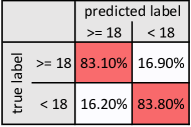
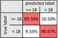
Figure 11 presents the confusion matrices for Group C: older/under 14 years old. Figure 11 LABEL:sub@fig:age_group_14 presents the detailed results for age group estimation avoiding the sex information. Figure 11 LABEL:sub@fig:age_group_14_F presents the detailed results for age group estimation on female individuals. Figure 11 LABEL:sub@fig:age_group_14_M presents the detailed results for age group estimation on male individuals.
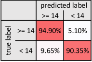
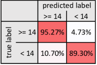
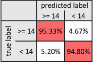
Figure 12 presents the confusion matrices with the detailed results for Group C: older/under 18 years old. Figure 12 LABEL:sub@fig:age_group_18 presents the detailed results for age group estimation avoiding the sex information. Figure 12 LABEL:sub@fig:age_group_18_F presents the detailed results for age group estimation on female individuals. Figure 12 LABEL:sub@fig:age_group_18_M presents the detailed results for age group estimation on male individuals.
IV Discussion
Estimating the age of victims and suspects of crimes is an essential procedure in the forensic casuistic of human identification [35, 36, 37, 38]. Age estimation becomes even more important when legal age thresholds are determined by the Court [39, 40]. Currently, the ages of 14 and 18 years represent the legal age threshold of sexual consent and majority in the judicial system of several countries worldwide [41, 42]. Designing scientific tools that allow the investigation of age with accuracy and evidence-based standards must be continuously encouraged to promote optimal forensic practices. Justified by the need for improving facial age estimation through photo-anthropometric analysis and founded on the hypothesis that the photo-anthropometric analyses of the human face can distinguish individuals younger and older than 14 and 18 years, this study aimed to propose and test an automatic solution to distinguish (male and female) individuals younger and older than 14 and 18 using photo-anthropometric analyses of the human face.
Population-specific studies on the use of photo-anthropometry of the face for age estimation are scarce in the scientific literature – especially those with large and standardized samples [9, 10, 11]. In this study, a large sample of Brazilian participants was collected (n=18000) and organized in a detailed, standardized data set available only for academic purposes. Sample standardization was accomplished by selecting male (n=500) and female (n=500) participants equally distributed (n=1000) in age intervals of one year (from 5 to 22 years). Moreover, all the digital photographs of the participants were taken with the same equipment and followed protocols previously reported in the scientific literature [9] and by the International Civil Aviation Organization (ICAO). In the scope of sampling strategy, an additional gain was obtained by choosing Brazilian participants, which are known for their multiracial formation [43] and phenotypic diversity [44]. This characteristic makes the study outcomes novel and eventually more reproducible in other populations.
In relation to the study outcomes, inferences about age were first investigated in association with sex. More specifically, the morphological information retrieved from the human face was tested based on its performance to classify males and females in age categories (5-22). A separate statistical test was evaluated for each of the 18 age groups. The outcomes endorsed the scientific literature by revealing difficulties for sexual dimorphism in young participants (12 years). In particular, the mean F1 scores in young age groups ranged between and . On the other hand, for participants aged from 13 to 22 years the mean F1 scores increased from to , showing evident improvement for classifying males and females. According to the scientific literature, the difference between young and old participants is explained by the lack of mature secondary sexual features depicted in facial photographs of children [45].
Recently, Kloess in [45] investigated the challenges in classifying child sexual abuse images. In their study, inferences about age (minor or not) were more easily and accurately given in images of babies and toddlers, while difficulties increased when the age of interest approached young adolescence. Interestingly, the larger size of the eyes in comparison with the other facial features and the presence of (milk) teeth and interdental gaps emerged as potential indicators of youth, while the use of make-up was a confounding factor [45]. In the present study, the diameter of the iris (iris ratio) [9] was used as fixed reference to calculate morphological ratios from the human face – which enables a quantification of the qualitative information provided by the previous authors. Additionally, photographs of participants depicting unnatural facial expressions (e.g. smiling) or using make-up were part of the exclusion criteria in the present study. Corroborated by the scientific literature [45], this methodological set up promoted a reduction of age classification bias as function of sex. In this context, sexual dimorphism, regardless of age, was performed as a quality-control procedure to eliminate the influence of age over the classification performance. The mean F1 score reached , indicating that the classification tool was able to properly distinguish most of the males and females of the sample if they were combined in a single group. In practice, the use of age-specific classification tools is recommended (whenever applicable) to best-fit the needs of each case – especially if the case involves age interests between 13 and 22 years, such as the age of sexual consent [42] and the age of legal majority [41].
In a second phase, the present study engaged in a deeper investigation based on age. This phase was justified by the uncertainty regarding the chronological age of victims and suspects of crimes that are commonly observed during the routine of forensic services. The methodological set up at this phase clustered together not only participants in age intervals of one year, but also in larger age intervals (e.g. within two, four and five years). Within each group, the performance was better for classifying the age of males. Between groups, the mean F1 scores were progressively higher with the increase in age interval size. Consequently, the best age estimates were found in the group with age interval of five years (mean F1 score: 0.74 combining males and females). Clearly, this outcome shows that the classification process becomes more difficult by refining the sample based on age. A similar approach was recently used by Machado in [9]. The authors performed a photo-anthropometric analysis to investigate the allometric growth of the human face by grouping together individuals within age intervals of four years. Despite the evident contributions towards the analysis of facial alterations over the time, the methodological set up proposed by the authors was limited compared to the present study. The advantages highlighted in the present setup include not only the sample stratification in groups of four different age intervals, but also the collection and quantification of much more morphological information from the human face. While the authors in [9] mapped human facial growth with ten measurements calculated from ratios based on the diameter of the iris, the present study outcomes were founded on 208 measures calculated with the same rationale. In practice, the improved methodological setup proposed in the present study induces more reliable and accurate age estimations.
The third and final phase addressed in this study focused on testing the classification system to distinguish participants that were younger or older than specific legal age thresholds of interest. This set up was justified to specifically meet the needs of justice when it comes to answer legal requests regarding the ages of 14 – related to sexual consent, and 18 – related to legal majority. The tests that were carried out in this phase showed, again, better classification of males, both in relation to the threshold of 14 and 18 years. When males and females were combined, the mean F1 scores reached 0.93 and 0.85 for the ages of 14 and 18 years, respectively. Satisfactory outcomes were also recently observed by Borges in [7] with a similar approach. The authors obtained accuracy (Area Under the Curve) estimates from Receiver Operating Characteristic (ROC) curve analyses that reached 0.96 and 0.90 for the ages of 14 and 18, respectively. Differences between studies include the larger sample size in the current investigation (n=18000 in face of 1000 used by the authors [7]) and, again, the larger number of measurements from the human face (n=208 compared to the 40 measurements used by the authors [7]). In forensic practice, the performance of the classification systems used in this study strengthens and supports its use for distinguishing victims and suspects of crimes aged below or above 14 and 18.
The automatic solution developed to classify individuals based on age and sex using morphological information retrieved from photo-anthropometric analyses of the human face reached optimal outcomes. More accurate age estimates were found in subjects aged between 13 and 22 years; estimates for the age of males were better than for females; and classifications based on legal age thresholds of sexual consent and majority were feasible and promising.
The proposed methodological setup presents multiple advantages compared to the available scientific literature. However, translating it to practice requires careful implementations and follow-up work to include updates in scientific evidence. Future studies in the field should test the reproducibility of this methodological setup and inherent outcomes in different populations. Investigations based on other legal age thresholds of interest are also encouraged to best-fit the judicial systems of different countries. Advances in the methodological setup for further improvements should include longitudinal sampling and three-dimensional imaging. Another approach in the computer vision field is to evaluate a classifier using deep learning techniques for age and sex estimation [46, 47], combining photos with photo-anthropometric indexes creating a cross-domain classifier. This allows the evaluation of possible improvements achieved by the inclusion of PAIs when compared to using only images as input.
Acknowledgment
The authors would like to acknowledge the team of Federal Police of Brazil, specially the forensic experts of National Institute of Criminalistic. This work was conducted with financial support from Coordination for the Improvement of Higher Education Personnel (CAPES) and Federal Police of Brazil (Grant Number: 001 Pro-Forenses 25/2014 CAPES).
References
- [1] N. Marquez-Grant, “An overview of age estimation in forensic anthropology: perspectives and practical considerations,” Annals of human biology, vol. 42, no. 4, pp. 308–322, 2015.
- [2] R. Silva, A. Franco, P. Dias, A. Gonçalves, and L. Paranhos, “Interrelationship between forensic radiology and forensic odontology—a case report of identified skeletal remains,” Journal of Forensic Radiology and Imaging, vol. 1, no. 4, pp. 201–206, 2013.
- [3] J. Adserias-Garriga, C. Thomas, D. H. Ubelaker, and S. C. Zapico, “When forensic odontology met biochemistry: Multidisciplinary approach in forensic human identification,” Archives of oral biology, vol. 87, pp. 7–14, 2018.
- [4] INTERPOL, “Interpol disaster victim identification guide,” https://www.interpol.int/INTERPOL-expertise/Forensics/DVI-Pages/DVI-guide, 2018. [Online]. Available: https://www.interpol.int/
- [5] C. Cattaneo, Z. Obertová, M. Ratnayake, L. Marasciuolo, J. Tutkuviene, P. Poppa, D. Gibelli, P. Gabriel, and S. Ritz-Timme, “Can facial proportions taken from images be of use for ageing in cases of suspected child pornography? a pilot study,” International journal of legal medicine, vol. 126, no. 1, pp. 139–144, 2012.
- [6] M. Ratnayake, Z. Obertová, M. Dose, P. Gabriel, H. Bröker, M. Brauckmann, A. Barkus, R. Rizgeliene, J. Tutkuviene, S. Ritz-Timme et al., “The juvenile face as a suitable age indicator in child pornography cases: a pilot study on the reliability of automated and visual estimation approaches,” International journal of legal medicine, vol. 128, no. 5, pp. 803–808, 2014.
- [7] D. L. Borges, F. B. Vidal, M. R. Flores, R. F. Melani, M. A. Guimarães, and C. E. Machado, “Photoanthropometric face iridial proportions for age estimation: An investigation using features selected via a joint mutual information criterion,” Forensic Science International, vol. 284, pp. 9 – 14, 2018.
- [8] C. Cattaneo, S. Ritz-Timme, P. Gabriel, D. Gibelli, E. Giudici, P. Poppa, D. Nohrden, S. Assmann, R. Schmitt, and M. Grandi, “The difficult issue of age assessment on pedo-pornographic material,” Forensic science international, vol. 183, no. 1, pp. e21–e24, 2009.
- [9] C. E. P. Machado, M. R. P. Flores, L. N. C. Lima, R. L. R. Tinoco, A. Franco, A. C. B. Bezerra, M. P. Evison, and M. A. Guimarães, “A new approach for the analysis of facial growth and age estimation: Iris ratio,” PLOS ONE, vol. 12, no. 7, p. e0180330, 2017.
- [10] M. R. Flores, C. E. Machado, M. D. Gallidabino, G. H. de Arruda, R. H. da Silva, F. B. de Vidal, and R. F. Melani, “Comparative assessment of a novel photo-anthropometric landmark-positioning approach for the analysis of facial structures on two-dimensional images,” Journal of forensic sciences, 2018.
- [11] P. S. Gonzales, C. E. P. Machado, and E. Michel-Crosato, “Photoanthropometry of the face in the young white brazilian population,” Brazilian dental journal, vol. 29, no. 6, pp. 619–623, 2018.
- [12] G. Zhu and S. van der Aa, “Trends of age of consent legislation in europe: A comparative study of 59 jurisdictions on the european continent,” New Journal of European Criminal Law, vol. 8, no. 1, pp. 14–42, 2017.
- [13] B. Carpenter, E. O’Brien, S. Hayes, and J. Death, “Harm, responsibility, age, and consent,” New Criminal Law Review: In International and Interdisciplinary Journal, vol. 17, no. 1, pp. 23–54, 2014.
- [14] G. O. Cericato, A. Franco, M. A. V. Bittencourt, M. A. P. Nunes, and L. R. Paranhos, “Correlating skeletal and dental developmental stages using radiographic parameters,” Journal of forensic and legal medicine, vol. 42, pp. 13–18, 2016.
- [15] M. A. Machado, E. D. Júnior, M. M. Fernandes, I. F. P. Lima, G. O. Cericato, A. Franco, and L. R. Paranhos, “Effectiveness of three age estimation methods based on dental and skeletal development in a sample of young brazilians,” Archives of oral biology, vol. 85, pp. 166–171, 2018.
- [16] International Organization for Standardization, “ISO/IEC 19794-5: Information technology – Biometric data interchange formats – Part 5: Face image data,” International Organization for Standardization, Standard, Mar. 2005.
- [17] M. R. Pinheiro-Flores and C. E. Palhares-Machado, Manual of facial photoanthropometry: landmarks in frontal view from visual references, 1st ed., 2017. [Online]. Available: http://facisgroup.org/facial_landmarks
- [18] M. R. Pinheiro-Flores, “Proposta de metodologia de análise fotoantropométrica para identificação humana em imagens faciais em norma frontal,” Master’s thesis, Faculdade de Odontologia de Ribeirão Preto, Universidade de São Paulo, 2014.
- [19] L. F. Porto, L. N. C. Lima, M. Flores, A. Valsecchi, O. Ibanez, C. E. M. Palhares, and F. de Barros Vidal. (2019, apr) Automatic cephalometric landmarks detection on frontal faces: an approach based on supervised learning techniques. [Online]. Available: https://arxiv.org/abs/1904.10816v1
- [20] J. Caple and C. Stephan, “A standardized nomenclature for craniofacial and facial anthropometry,” International Journal of Legal Medicine, vol. 130, no. 3, pp. 863–879, 2016.
- [21] R. E. Brown, T. P. Kelliher, P. H. Tu, W. D. Turner, M. A. Taister, and K. W. Miller, “A survey of tissue-depth landmarks for facial approximation,” Forensic Sci. Commun, vol. 6, no. 1, 2004.
- [22] P. J. Phillips, H. Moon, S. Rizvi, P. J. Rauss et al., “The feret evaluation methodology for face-recognition algorithms,” Pattern Analysis and Machine Intelligence, IEEE Transactions on, vol. 22, no. 10, pp. 1090–1104, 2000.
- [23] S. Apeland, “Intel ai devcloud,” https://www.intel.ai/devcloud/, accessed: 2019-01-22. [Online]. Available: https://www.intel.ai/devcloud/
- [24] F. Chollet et al., “Keras,” https://keras.io, 2015.
- [25] M. Abadi, A. Agarwal, P. Barham, E. Brevdo, Z. Chen, C. Citro, G. S. Corrado, A. Davis, J. Dean, M. Devin, S. Ghemawat, I. Goodfellow, A. Harp, G. Irving, M. Isard, Y. Jia, R. Jozefowicz, L. Kaiser, M. Kudlur, J. Levenberg, D. Mané, R. Monga, S. Moore, D. Murray, C. Olah, M. Schuster, J. Shlens, B. Steiner, I. Sutskever, K. Talwar, P. Tucker, V. Vanhoucke, V. Vasudevan, F. Viégas, O. Vinyals, P. Warden, M. Wattenberg, M. Wicke, Y. Yu, and X. Zheng, “TensorFlow: Large-scale machine learning on heterogeneous systems,” 2015, software available from tensorflow.org. [Online]. Available: https://www.tensorflow.org/
- [26] M. W. Gardner and S. Dorling, “Artificial neural networks (the multilayer perceptron)—a review of applications in the atmospheric sciences,” Atmospheric environment, vol. 32, no. 14-15, pp. 2627–2636, 1998.
- [27] D. P. Kingma and J. Ba, “Adam: A method for stochastic optimization,” arXiv preprint arXiv:1412.6980, 2014.
- [28] D. M. W. Powers, “Evaluation: From precision, recall and f-measure to roc., informedness, markedness & correlation,” Journal of Machine Learning Technologies, vol. 2, no. 1, pp. 37–63, 2011.
- [29] F. Provost and R. Kohavi, “On applied research in machine learning,” in Machine learning, 1998, pp. 127–132.
- [30] T. Hastie, R. Tibshirani, and J. Friedman, The elements of statistical learning: data mining, inference and prediction, 2nd ed. Springer, 2009. [Online]. Available: http://www-stat.stanford.edu/ tibs/ElemStatLearn/
- [31] R. Kohavi et al., “A study of cross-validation and bootstrap for accuracy estimation and model selection,” in Ijcai, vol. 14. Stanford, CA, 1995, pp. 1137–1145.
- [32] I. Goodfellow, J. Pouget-Abadie, M. Mirza, B. Xu, D. Warde-Farley, S. Ozair, A. Courville, and Y. Bengio, “Generative adversarial nets,” in Advances in Neural Information Processing Systems 27, Z. Ghahramani, M. Welling, C. Cortes, N. D. Lawrence, and K. Q. Weinberger, Eds. Curran Associates, Inc., 2014, pp. 2672–2680. [Online]. Available: http://papers.nips.cc/paper/5423-generative-adversarial-nets.pdf
- [33] M. B. WILK and S. S. SHAPIRO, “An analysis of variance test for normality (complete samples),” Biometrika, vol. 52, no. 3-4, pp. 591–611, 12 1965. [Online]. Available: https://doi.org/10.1093/biomet/52.3-4.591
- [34] M. Kutner, Applied Linear Statistical Models, ser. McGrwa-Hill international edition. McGraw-Hill Irwin, 2005. [Online]. Available: https://books.google.com.br/books?id=0xqCAAAACAAJ
- [35] A. Schmeling, A. Olze, W. Reisinger, and G. Geserick, “Age estimation of living people undergoing criminal proceedings,” The Lancet, vol. 358, no. 9276, pp. 89–90, 2001.
- [36] A. Schmeling, R. Dettmeyer, E. Rudolf, V. Vieth, and G. Geserick, “Forensic age estimation: methods, certainty, and the law,” Deutsches Ärzteblatt International, vol. 113, no. 4, p. 44, 2016.
- [37] R. F. Silva, S. D. S. C. Mendes, A. F. do Rosário Júnior, P. E. M. Dias, and L. B. Martorell, “Evidência documental x evidência biológica para estimativa da idade–relato de caso pericial.” Revista Odontológica do Brasil Central, vol. 22, no. 60, 2013.
- [38] A. L. R. Machado, T. U. Dezem, A. T. Bruni, and R. H. A. da Silva, “Age estimation by facial analysis based on applications available for smartphones,” The Journal of forensic odonto-stomatology, vol. 35, no. 2, p. 55, 2017.
- [39] A. R. Deitos, C. Costa, E. Michel-Crosato, I. Galić, R. Cameriere, and M. G. H. Biazevic, “Age estimation among brazilians: younger or older than 18?” Journal of forensic and legal medicine, vol. 33, pp. 111–115, 2015.
- [40] B. M. Santiago, L. Almeida, Y. W. Cavalcanti, M. B. Magno, and L. C. Maia, “Accuracy of the third molar maturity index in assessing the legal age of 18 years: a systematic review and meta-analysis,” International journal of legal medicine, vol. 132, no. 4, pp. 1167–1184, 2018.
- [41] A. Franco, P. Thevissen, S. Fieuws, P. H. C. Souza, and G. Willems, “Applicability of willems model for dental age estimations in brazilian children,” Forensic science international, vol. 231, no. 1-3, pp. 401–e1, 2013.
- [42] H. Graupner, “Sexual consent: The criminal law in europe and overseas,” Archives of Sexual Behavior, vol. 29, no. 5, pp. 415–461, 2000.
- [43] V. S. d. Souza, “Science and miscegenation in the early twentieth century: Edgard roquette-pinto’s debates and controversies with us physical anthropology,” História, Ciências, Saúde-Manguinhos, vol. 23, no. 3, pp. 597–614, 2016.
- [44] A. Edmonds, “Triumphant miscegenation: Reflections on beauty and race in brazil,” Journal of Intercultural Studies, vol. 28, no. 1, pp. 83–97, 2007.
- [45] J. A. Kloess, J. Woodhams, H. Whittle, T. Grant, and C. E. Hamilton-Giachritsis, “The challenges of identifying and classifying child sexual abuse material,” Sexual Abuse, p. 1079063217724768, 2017.
- [46] Q. Cao, L. Shen, W. Xie, O. M. Parkhi, and A. Zisserman, “Vggface2: A dataset for recognising faces across pose and age,” in 2018 13th IEEE International Conference on Automatic Face & Gesture Recognition (FG 2018). IEEE, 2018, pp. 67–74.
- [47] J. Xing, K. Li, W. Hu, C. Yuan, and H. Ling, “Diagnosing deep learning models for high accuracy age estimation from a single image,” Pattern Recognition, vol. 66, pp. 106–116, 2017.
Appendix A Confusion Matrices
Appendix B Photo-anthropometric Indexes
| PAI | Landmarks | Description |
|---|---|---|
| PAI-0 | al_r-al_l al_l-al_r | Nose width |
| PAI-1 | al_r-ch_r al_l-ch_l | Wing of the nose - Labial Commissure (Same side) |
| PAI-2 | al_r-ch_l al_l-ch_r | Wing of the nose - Labial Commissure (Different side) |
| PAI-3 | al_r-cph_r al_l-cph_l | Wing of the nose - Crista philtri (Same side) |
| PAI-4 | al_r-cph_l al_l-cph_r | Wing of the nose - Crista philtri (Different side) |
| PAI-5 | al_r-ec_r al_l-ec_l | Wing of the nose - Ectocanthion (Same side) |
| PAI-6 | al_r-ec_l al_l-ec_r | Wing of the nose - Ectocanthion (Different side) |
| PAI-7 | al_r-en_r al_l-en_l | Wing of the nose - Endocanthion (Same side) |
| PAI-8 | al_r-en_l al_l-en_r | Wing of the nose - Endocanthion (Different side) |
| PAI-9 | al_r-g al_l-g | Wing of the nose - Glabella |
| PAI-10 | al_r-gn al_l-gn | Wing of the nose - Chin |
| PAI-11 | al_r-go_r al_l-go_l | Wing of the nose - Gonion (Same side) |
| PAI-12 | al_r-go_l al_l-go_r | Wing of the nose - Gonion (Different side) |
| PAI-13 | al_r-il_r al_l-il_l | Wing of the nose - Lateral iris (Same side) |
| PAI-14 | al_r-il_l al_l-il_r | Wing of the nose - Lateral iris (Different side) |
| PAI-15 | al_r-im_r al_l-im_l | Wing of the nose - Medial iris (Same side) |
| PAI-16 | al_r-im_l al_l-im_r | Wing of the nose - Medial iris (Different side) |
| PAI-17 | al_r-li al_l-li | Wing of the nose - Lower lip |
| PAI-18 | al_r-ls al_l-ls | Wing of the nose - Upper lip |
| PAI-19 | al_r-mid al_l-mid | Wing of the nose - Midnasal |
| PAI-20 | al_r-n al_l-n | Wing of the nose - Nasion |
| PAI-21 | al_r-pu_r al_l-pu_l | Wing of the nose - Pupil (Same side) |
| PAI-22 | al_r-pu_l al_l-pu_r | Wing of the nose - Pupil (Different side) |
| PAI-23 | al_r-sn al_l-sn | Wing of the nose - Base of the nose |
| PAI-24 | al_r-sto al_l-sto | Wing of the nose - Stomion |
| PAI-25 | al_r-zy_r al_l-zy_l | Wing of the nose - Zygion (Same side) |
| PAI-26 | al_r-zy_l al_l-zy_r | Wing of the nose - Zygion (Different side) |
| PAI-27 | ch_r-ch_l ch_l-ch_r | Mouth width |
| PAI-28 | ch_r-cph_r ch_l-cph_l | Labial Commissure - Crista philtri (Same side) |
| PAI-29 | ch_r-cph_l ch_l-cph_r | Labial Commissure - Crista philtri (Different side) |
| PAI-30 | ch_r-ec_r ch_l-ec_l | Labial Commissure - Ectocanthion (Same side) |
| PAI-31 | ch_r-ec_l ch_l-ec_r | Labial Commissure - Ectocanthion (Different side) |
| PAI-32 | ch_r-en_r ch_l-en_l | Labial Commissure - Endocanthion (Same side) |
| PAI-33 | ch_r-en_l ch_l-en_r | Labial Commissure - Endocanthion (Different side) |
| PAI-34 | ch_r-g ch_l-g | Labial Commissure - Glabella |
| PAI-35 | ch_r-gn ch_l-gn | Labial Commissure - Chin |
| PAI-36 | ch_r-go_r ch_l-go_l | Labial Commissure - Gonion (Same side) |
| PAI-37 | ch_r-go_l ch_l-go_r | Labial Commissure - Gonion (Different side) |
| PAI-38 | ch_r-il_r ch_l-il_l | Labial Commissure - Lateral iris (Same side) |
| PAI-39 | ch_r-il_l ch_l-il_r | Labial Commissure - Lateral iris (Different side) |
| PAI-40 | ch_r-im_r ch_l-im_l | Labial Commissure - Medial iris (Same side) |
| PAI-41 | ch_r-im_l ch_l-im_r | Labial Commissure - Medial iris (Different side) |
| PAI-42 | ch_r-li ch_l-li | Labial Commissure - Lower lip |
| PAI-43 | ch_r-ls ch_l-ls | Labial Commissure - Upper lip |
| PAI-44 | ch_r-mid ch_l-mid | Labial Commissure - Midnasal |
| PAI-45 | ch_r-n ch_l-n | Labial Commissure - Nasion |
| PAI-46 | ch_r-pu_r ch_l-pu_l | Labial Commissure - Pupil (Same side) |
| PAI-47 | ch_r-pu_l ch_l-pu_r | Labial Commissure - Pupil (Different side) |
| PAI-48 | ch_r-sn ch_l-sn | Labial Commissure - Base of the nose |
| PAI-49 | ch_r-sto ch_l-sto | Labial Commissure - Stomion |
| PAI-50 | ch_r-zy_r ch_l-zy_l | Labial Commissure - Zygion (Same side) |
| PAI-51 | ch_r-zy_l ch_l-zy_r | Labial Commissure - Zygion (Different side) |
| PAI-52 | cph_r-cph_l cph_l-cph_r | Width of the crista philtri |
| PAI-53 | cph_r-ec_r cph_l-ec_l | Crista philtri - Ectocanthion (Same side) |
| PAI-54 | cph_r-ec_l cph_l-ec_r | Crista philtri - Ectocanthion (Different side) |
| PAI-55 | cph_r-en_r cph_l-en_l | Crista philtri - Endocanthion (Same side) |
| PAI-56 | cph_r-en_l cph_l-en_r | Crista philtri - Endocanthion (Different side) |
| PAI-57 | cph_r-g cph_l-g | Crista philtri - Glabella |
| PAI-58 | cph_r-gn cph_l-gn | Crista philtri - Chin |
| PAI-59 | cph_r-go_r cph_l-go_l | Crista philtri - Gonion (Same side) |
| PAI-60 | cph_r-go_l cph_l-go_r | Crista philtri - Gonion (Different side) |
| PAI-61 | cph_r-il_r cph_l-il_l | Crista philtri - Lateral iris (Same side) |
| PAI-62 | cph_r-il_l cph_l-il_r | Crista philtri - Lateral iris (Different side) |
| PAI-63 | cph_r-im_r cph_l-im_l | Crista philtri - Medial iris (Same side) |
| PAI-64 | cph_r-im_l cph_l-im_r | Crista philtri - Medial iris (Different side) |
| PAI-65 | cph_r-li cph_l-li | Crista philtri - Lower lip |
| PAI-66 | cph_r-ls cph_l-ls | Crista philtri - Upper lip |
| PAI-67 | cph_r-mid cph_l-mid | Crista philtri - Midnasal |
| PAI-68 | cph_r-n cph_l-n | Crista philtri - Nasion |
| PAI-69 | cph_r-pu_r cph_l-pu_l | Crista philtri - Pupil (Same side) |
| PAI-70 | cph_r-pu_l cph_l-pu_r | Crista philtri - Pupil (Different side) |
| PAI-71 | cph_r-sn cph_l-sn | Crista philtri - Base of the nose |
| PAI-72 | cph_r-sto cph_l-sto | Crista philtri - Stomion |
| PAI-73 | cph_r-zy_r cph_l-zy_l | Crista philtri - Zygion (Same side) |
| PAI-74 | cph_r-zy_l cph_l-zy_r | Crista philtri - Zygion (Different side) |
| PAI-75 | ec_r-ec_l ec_l-ec_r | Width of the ectocanthion |
| PAI-76 | ec_r-en_r ec_l-en_l | Eye width |
| PAI-77 | ec_r-en_l ec_l-en_r | Ectocanthion - Endocanthion (Different side) |
| PAI-78 | ec_r-g ec_l-g | Ectocanthion - Glabella |
| PAI-79 | ec_r-gn ec_l-gn | Ectocanthion - Chin |
| PAI-80 | ec_r-go_r ec_l-go_l | Ectocanthion - Gonion (Same side) |
| PAI-81 | ec_r-go_l ec_l-go_r | Ectocanthion - Gonion (Different side) |
| PAI-82 | ec_r-il_r ec_l-il_l | Ectocanthion - Lateral iris (Same side) |
| PAI-83 | ec_r-il_l ec_l-il_r | Ectocanthion - Lateral iris (Different side) |
| PAI-84 | ec_r-im_r ec_l-im_l | Ectocanthion - Medial iris (Same side) |
| PAI-85 | ec_r-im_l ec_l-im_r | Ectocanthion - Medial iris (Different side) |
| PAI-86 | ec_r-li ec_l-li | Ectocanthion - Lower lip |
| PAI-87 | ec_r-ls ec_l-ls | Ectocanthion - Upper lip |
| PAI-88 | ec_r-mid ec_l-mid | Ectocanthion - Midnasal |
| PAI-89 | ec_r-n ec_l-n | Ectocanthion - Nasion |
| PAI-90 | ec_r-pu_r ec_l-pu_l | Ectocanthion - Pupil (Same side) |
| PAI-91 | ec_r-pu_l ec_l-pu_r | Ectocanthion - Pupil (Different side) |
| PAI-92 | ec_r-sn ec_l-sn | Ectocanthion - Base of the nose |
| PAI-93 | ec_r-sto ec_l-sto | Ectocanthion - Stomion |
| PAI-94 | ec_r-zy_r ec_l-zy_l | Ectocanthion - Zygion (Same side) |
| PAI-95 | ec_r-zy_l ec_l-zy_r | Ectocanthion - Zygion (Different side) |
| PAI-96 | en_r-en_l en_l-en_r | Inter-canthion width |
| PAI-97 | en_r-g en_l-g | Endocanthion - Glabella |
| PAI-98 | en_r-gn en_l-gn | Endocanthion - Chin |
| PAI-99 | en_r-go_r en_l-go_l | Endocanthion - Gonion (Same side) |
| PAI-100 | en_r-go_l en_l-go_r | Endocanthion - Gonion (Different side) |
| PAI-101 | en_r-il_r en_l-il_l | Endocanthion - Lateral iris (Same side) |
| PAI-102 | en_r-il_l en_l-il_r | Endocanthion - Lateral iris (Different side) |
| PAI-103 | en_r-im_r en_l-im_l | Endocanthion - Medial iris (Same side) |
| PAI-104 | en_r-im_l en_l-im_r | Endocanthion - Medial iris (Different side) |
| PAI-105 | en_r-li en_l-li | Endocanthion - Lower lip |
| PAI-106 | en_r-ls en_l-ls | Endocanthion - Upper lip |
| PAI-107 | en_r-mid en_l-mid | Endocanthion - Midnasal |
| PAI-108 | en_r-n en_l-n | Endocanthion - Nasion |
| PAI-109 | en_r-pu_r en_l-pu_l | Endocanthion - Pupil (Same side) |
| PAI-110 | en_r-pu_l en_l-pu_r | Endocanthion - Pupil (Different side) |
| PAI-111 | en_r-sn en_l-sn | Endocanthion - Base of the nose |
| PAI-112 | en_r-sto en_l-sto | Endocanthion - Stomion |
| PAI-113 | en_r-zy_r en_l-zy_l | Endocanthion - Zygion (Same side) |
| PAI-114 | en_r-zy_l en_l-zy_r | Endocanthion - Zygion (Different side) |
| PAI-115 | g-gn | Height of the face |
| PAI-116 | g-go_r g-go_l | Glabella - Gonion (Same side) |
| PAI-117 | g-il_r g-il_l | Glabella - Gonion (Different side) |
| PAI-118 | g-im_r g-im_l | Glabella - Medial iris |
| PAI-119 | g-li | Glabella - Upper lip |
| PAI-120 | g-ls | Glabella - Lower lip |
| PAI-121 | g-mid | Glabella Midnasal |
| PAI-122 | g-n | Glabella - Nasion |
| PAI-123 | g-pu_r g-pu_l | Glabella - Pupil |
| PAI-124 | g-sn | Glabella - Base of the nose |
| PAI-125 | g-sto | Glabella - Stomion |
| PAI-126 | g-zy_r g-zy_l | Glabella - Zygion |
| PAI-127 | gn-go_r gn-go_l | Chin - Gonion |
| PAI-128 | gn-il_r gn-il_l | Chin - Lateral iris |
| PAI-129 | gn-im_r gn-im_l | Chin - Medial iris |
| PAI-130 | gn-li | Chin - Lower lip |
| PAI-131 | gn-ls | Chin - Upper lip |
| PAI-132 | gn-mid | Chin - Midnasal |
| PAI-133 | gn-n | Chin - Nasion |
| PAI-134 | gn-pu_r gn-pu_l | Chin - Pupil |
| PAI-135 | gn-sn | Chin - Base of the nose |
| PAI-136 | gn-sto | Chin - Stomion |
| PAI-137 | gn-zy_r gn-zy_l | Chin - Zygion |
| PAI-138 | go_r-go_l go_l-go_r | Inter-gonion width |
| PAI-139 | go_r-il_r go_l-il_l | Gonion - Lateral iris (Same side) |
| PAI-140 | go_r-il_l go_l-il_r | Gonion - Lateral iris (Different side) |
| PAI-141 | go_r-im_r go_l-im_l | Gonion - Medial iris (Same side) |
| PAI-142 | go_r-im_l go_l-im_r | Gonion - Medial iris (Different side) |
| PAI-143 | go_r-li go_l-li | Gonion - Lower lip |
| PAI-144 | go_r-ls go_l-ls | Gonion - Upper lip |
| PAI-145 | go_r-mid go_l-mid | Gonion - Midnasal |
| PAI-146 | go_r-n go_l-n | Gonion - Nasion |
| PAI-147 | go_r-pu_r go_l-pu_l | Gonion - Pupil (Same side) |
| PAI-148 | go_r-pu_l go_l-pu_r | Gonion - Pupil (Different side) |
| PAI-149 | go_r-sn go_l-sn | Gonion - Base of the nose |
| PAI-150 | go_r-sto go_l-sto | Gonion - Stomion |
| PAI-151 | go_r-zy_r go_l-zy_l | Gonion - Zygion (Same side) |
| PAI-152 | go_r-zy_l go_l-zy_r | Gonion - Zygion (Different side) |
| PAI-153 | il_r-il_l il_l-il_r | Maximum iris width |
| PAI-154 | il_r-im_r il_l-im_l | Diameter of the iris |
| PAI-155 | il_r-im_l il_l-im_r | Lateral iris - Medial iris (Different side) |
| PAI-156 | il_r-li il_l-li | Lateral iris - Upper lip |
| PAI-157 | il_r-ls il_l-ls | Lateral iris - Upper lip |
| PAI-158 | il_r-mid il_l-mid | Lateral iris - Midnasal |
| PAI-159 | il_r-n il_l-n | Lateral iris - Nasion |
| PAI-160 | il_r-pu_r il_l-pu_l | Lateral iris - Pupil (Same side) |
| PAI-161 | il_r-pu_l il_l-pu_r | Lateral iris - Pupil (Different side) |
| PAI-162 | il_r-sn il_l-sn | Lateral iris - Base of the nose |
| PAI-163 | il_r-sto il_l-sto | Lateral iris - Stomion |
| PAI-164 | il_r-zy_r il_l-zy_l | Lateral iris - Zygion (Same side) |
| PAI-165 | il_r-zy_l il_l-zy_r | Lateral iris - Zygion (Different side) |
| PAI-166 | im_r-im_l im_l-im_r | Minimum iris width |
| PAI-167 | im_r-li im_l-li | Medial iris - Lower lip |
| PAI-168 | im_r-ls im_l-ls | Medial iris - Upper lip |
| PAI-169 | im_r-mid im_l-mid | Medial iris - Midnasal |
| PAI-170 | im_r-n im_l-n | Medial iris - Nasion |
| PAI-171 | im_r-pu_r im_l-pu_l | Medial iris - Pupil (Same side) |
| PAI-172 | im_r-pu_l im_l-pu_r | Medial iris - Pupil (Different side) |
| PAI-173 | im_r-sn im_l-sn | Medial iris - Base of the nose |
| PAI-174 | im_r-sto im_l-sto | Medial iris - Stomion |
| PAI-175 | im_r-zy_r im_l-zy_l | Medial iris - Zygion (Same side) |
| PAI-176 | im_r-zy_l im_l-zy_r | Medial iris - Zygion (Different side) |
| PAI-177 | li-ls | Lip height |
| PAI-178 | li-mid | Lower lip - Midnasal |
| PAI-179 | li-n | Lower lip - Nasion |
| PAI-180 | li-pu_r li-pu_l | Lower lip - Pupil |
| PAI-181 | li-sn | Lower lip - Base of the nose |
| PAI-182 | li-sto | Lower lip - Stomion |
| PAI-183 | li-zy_r li-zy_l | Lower lip - Zygion |
| PAI-184 | ls-mid | Upper lip - Midnasal |
| PAI-185 | ls-n | Upper lip - Nasion |
| PAI-186 | ls-pu_r ls-pu_l | Upper lip - Pupil |
| PAI-187 | ls-sn | Upper lip - Base of the nose |
| PAI-188 | ls-sto | Upper lip - Stomion |
| PAI-189 | ls-zy_r ls-zy_l | Upper lip - Zygion |
| PAI-190 | mid-n | Midnasal - Nasion |
| PAI-191 | mid-pu_r mid-pu_l | Midnasal - Pupil |
| PAI-192 | mid-sn | Midnasal - Base of the nose |
| PAI-193 | mid-sto | Midnasal - Stomion |
| PAI-194 | mid-zy_r mid-zy_l | Midnasal - Zygion |
| PAI-195 | n-pu_r n-pu_l | Nasion - Pupil |
| PAI-196 | n-sn | Nose height |
| PAI-197 | n-sto | Nasion - Stomion |
| PAI-198 | n-zy_r n-zy_l | Nasion - Zygion |
| PAI-199 | pu_r-pu_l pu_l-pu_r | Inter-pupil width |
| PAI-200 | pu_r-sn pu_l-sn | Pupil - Base of the nose |
| PAI-201 | pu_r-sto pu_l-sto | Pupil - Stomion |
| PAI-202 | pu_r-zy_r pu_l-zy_l | Pupil - Zygion (Same side) |
| PAI-203 | pu_r-zy_l pu_l-zy_r | Pupil - Zygion (Different side) |
| PAI-204 | sn-sto | Base of the nose - Stomion |
| PAI-205 | sn-zy_r sn-zy_l | Base of the nose - Zygion |
| PAI-206 | sto-zy_r sto-zy_l | Stomion - Zygion |
| PAI-207 | zy_r-zy_l zy_l-zy_r | Face width |