CAD
short=CAD,
long=computer-aided diagnosis
\DeclareAcronymPXR
short=PXR,
long=pelvic X-ray
\DeclareAcronymCXR
short=CXR,
long=chest X-ray
\DeclareAcronymCNN
short=CNN,
long=convolutional neural network
\DeclareAcronymAUC
short=AUCROC,
long=area under the ROC curve,
long-plural-form=areas under the ROC curve
\DeclareAcronymROC
short=ROC,
long=receiver operating characteristic
\DeclareAcronymROI
short=ROI,
long=region of interest
\DeclareAcronymFCN
short=FCN,
long=fully-convolutional network
\DeclareAcronymMIL
short=MIL,
long=multiple instance learning
\DeclareAcronymPACS
short=PACS,
long=picture archiving and communication system
\DeclareAcronymER
short=ER,
long=emergency room
\DeclareAcronymSR
short=S@R,
long=specificity at recall rate of
\DeclareAcronymRS
short=R@S,
long=recall at specificity rate of
\DeclareAcronymPR
short=P@R,
long=precision at recall rate of
\DeclareAcronymLSE
short=LSE,
long=log of the sum of the exponentials
\DeclareAcronymBCE
short=BCE,
long=binary cross entropy
\DeclareAcronymGAP
short=GAP,
long=global average pooling
11institutetext: 1PAII Inc., Bethesda, MD, USA
2Chang Gung Memorial Hospital, Linkou, Taiwan, ROC
3Ping An Technology, Shenzhen, China
Weakly Supervised Universal Fracture Detection in Pelvic X-rays
Abstract
Hip and pelvic fractures are serious injuries with life-threatening complications. However, diagnostic errors of fractures in \acpPXR are very common, driving the demand for \acCAD solutions. A major challenge lies in the fact that fractures are localized patterns that require localized analyses. Unfortunately, the \acpPXR residing in hospital \aclPACS do not typically specify \aclpROI. In this paper, we propose a two-stage hip and pelvic fracture detection method that executes localized fracture classification using weakly supervised \acROI mining. The first stage uses a large capacity \aclFCN, i.e., deep with high levels of abstraction, in a \aclMIL setting to automatically mine probable true positive and definite hard negative \acpROI from the whole \acPXR in the training data. The second stage trains a smaller capacity model, i.e., shallower and more generalizable, with the mined \acpROI to perform localized analyses to classify fractures. During inference, our method detects hip and pelvic fractures in one pass by chaining the probability outputs of the two stages together. We evaluate our method on \acpPXR, reporting an \aclAUC value of , the highest among state-of-the-art fracture detection methods. Moreover, we show that our two-stage approach can perform comparably to human physicians (even outperforming emergency physicians and surgeons), in a preliminary reader study of readers.
Keywords:
Fracture classification and localization, Pelvic X-ray, Weakly supervised detection, Cascade two-stage training, Image level labels
1 Introduction
Hip and pelvic fractures belong to a frequent trauma injury category worldwide [8]. Frontal \acpPXR are the standard imaging tool for diagnosing pelvic and hip fractures in the \acER. However, anatomical complexities and perspective projection distortions contribute to a high rate of diagnostic errors [2] that may delay treatment and increase patient care cost, morbidity, and mortality [10]. As such, an effective \acPXR \acCAD approach for both pelvic and hip fractures is of high clinical interest, with the aim of reducing diagnostic errors and improving patient outcomes.
Image-level labels are the only supervisory signal typically available in \acPACS data. Thus, a widely adopted formulation for X-ray abnormality detection is a single-stage global classifier [1, 11, 9, 3]. However, for \acpPXR this approach is challenged by the localized nature of fractures and the complexity of the surrounding anatomical regions. Moreover, such global classifiers can be prone to overfitting, as it is unlikely that a training dataset could capture the combinatorial complexity of configurations of fracture locations, orientations, and background contexts within the whole \acPXR—this complexity is analogous to similar challenges within computer vision [13]. Indeed, for hip fractures alone, Jiménez-Sánchez et al. show that using localized \acpROI produces significantly better F1 scores over a global approach [7] and Gale et al. achieve impressive \acpAUC of by first automatically extracting \acpROI centered on the femoral neck [4]. These recent results bolster the argument for concentrating on local fracture patterns.
Nonetheless, the above prior work all only focuses on diagnosing hip fractures and does not attempt to classify the more complex pelvic fractures (fractures in three pelvic bones: the ilium, ischium, and pubis). As Fig. 1 illustrates, the makeup of pelvis fractures is much more complex, as there are a large variety of possible types with very different visual patterns at various locations. In addition, pelvic bones overlap with the lower abdomen, further confounding image patterns. Finally, unlike hip fractures, which occur at the femoral neck/head, pelvic fractures can occur anywhere on the large pelvis, both increasing the aforementioned image pattern combinatorial complexity and precluding automatic \acROI extraction based on anatomy alone, such as was done in prior work [4]. Thus, while using \acROI-based classification is even more desirable for pelvic fractures, it is paradoxically more challenging to extract said \acpROI.
To bridge this gap, we propose a two-stage weakly supervised \acROI mining and subsequent classification method for \acPXR fracture classification. In the first stage, we train a weakly-supervised, but high capacity, \acMIL \acFCN to mine local probable positive and definite hard negative \acpROI. In the second stage, we use the mined \acpROI to train a lower capacity network in a fully-supervised setting. During inference, the two networks are chained together to provide a complete classification solution. Experiments use a dataset of \acpPXR, with only image-level labels, that we collected from the \acPACS of Chang Gung Memorial Hospital. We show that single-stage classifiers, whether low- or high-capacity, are unable to match our two-stage approach. Our chained two-stage method outperforms the best single-stage alternative, with a \acSR of compared to , and corresponding improvements in \acpAUC. Moreover, using an independent reader study of patients, our system achieves an accuracy of , which is equivalent to physicians. As such, we are the first to tackle automatic \acPXR pelvis fracture classification and also the first to demonstrate diagnostic performance equivalent to human physicians for both hip and pelvic fractures.
2 Method
Fig. 2 depicts the overall workflow of our chained two-stage pelvic and hip fracture detection method. We elaborate on the two stages below.
2.1 Weakly-Supervised \acsROI Mining
In the first stage, we train an \acFCN using a deep \acMIL formulation [12], employing the large-capacity DenseNet-121 [6] network as backbone. The DenseNet-121 features are then processed using a convolutional layer and a sigmoid activation to produce a probability map. Owing to the localized properties of \acpFCN, each value of the probability map can be interpreted as the probability of fracture in the corresponding region in the input \acPXR. The maximum value would then represent the probability of fracture within the entire image. Instead, we use \acLSE pooling, which is a differentiable approximation of max pooling, given by
| (1) |
where is the probability map, and is a hyper-parameter controlling the behavior of \acLSE between max pooling () and average pooling (). With the pooled global probability, \acBCE loss is calculated against the image level label, and is used to train the network. While, this formulation has been applied directly for weakly supervised abnormality detection in \acpCXR [12], as we show in our results this approach’s performance is limited for hip and pelvic fracture detection. Therefore, we use the \acFCN as a proposal generator to mine \acpROI from the training data.
To mine \acpROI from the training data to train a localized classification model, we first create an image-level classifier using , and select a threshold corresponding to a high sensitivity on the training data (we use in our experiments). We then extract up to \acpROI from each \acPXR in the training data in every training epoch of the second stage model. Specifically, for \acpPXR with positive ground-truth image-level labels, i.e., with fracture(s), up to locations are randomly selected from
| (2) |
These \acpROI are labeled as probable fracture positive. For \acpPXR with negative ground-truth image-level labels, i.e., no fractures, the same \acROI extraction strategy selects up to \acpROI. These are considered as definite hard negatives. If there are less than hard negatives extracted, additional negative \acpROI are randomly extracted from the \acPXR to make up the total. The \acpROI produced using the above strategy contains probable positives, hard negatives, and easy negatives. Although this approach adds a degree of label noise due to the probable positive \acsROI, as we outline in the following, this comes with the added benefits of using a subsequent localized and more generalizable \acROI classifier.
2.2 Fracture \acsROI Classification
In the second stage, we use the \acpROI mined from the first stage as training data for a fully supervised localized classification network. Since the positive samples are mostly \acpROI around fractures with limited background context, the visual patterns of fractures become more dominant, simplifying the classification task. In addition, the distribution of mined \acpROI are heavily weighted toward hard negatives, i.e., false positive regions from the first stage. This concentrates the modeling power of the second-stage classifier on differentiating these difficult/confusing fracture-like patterns. As a result, we are able to train a smaller capacity network, e.g., ResNet-18 [5], to reliably classify the \acpROI, which is more generalizable and less prone to overfitting compared to a high-capacity network modeling the entire \acPXR.
During inference, the two stages are chained together to provide a complete solution. The first stage \acFCN acts as a proposal generator, and the highest value from the probability map is selected, denoted as , along with the corresponding \acROI. The second stage classifier is then applied on the proposed \acROI to produce a fracture probability score, denoted . The final image-level probability of fracture is computed by multiplying the two probability scores, . As such, we use the second stage classifier as a filter to reject false positives from the first stage.
Hip/pelvic fracture differentiation: Our two-stage method detects hip and pelvic fractures as one class, because the most important goal of \acPXR \acCAD is to detect fractures. Using one universal fracture class also helps to prevent the model from picking up co-occurrence relationships between hip/pelvic fractures that may be overly represented from the current training data [13]. In scenarios where hip and pelvic fractures do need to be differentiated, e.g., automatic medical image reporting, an additional classification output node can be added to the second stage model. Similar to the hierarchical classification schemes [3], the new node is trained only on positive fracture \acpROI mined in the first stage. Like fracture classification, during inference hip/pelvic fracture differentiation can be obtained in one feed-forward pass of the network.
3 Experiments and Results
We evaluate our framework using \acPXR images collected from the \acPACS of Chang Gung Memorial Hospital, corresponding to patients in the trauma registry. We resized all images to pixels. The final dataset consisted of images, including images with fractures ( and hip and pelvic fractures, respectively). Besides this dataset, we also collected an independent \acPXR dataset, containing cases ( hip fractures, pelvic fractures, and no findings) for a reader study comparing our approach with that of physicians.
We use ImageNet pre-trained weights to initialize the networks in both stages. The Adam optimizer was used to train both models for epochs with a batch size of and a starting learning rate of reduced by a factor of upon plateaus. In addition to \acAUC, we measure \acfSR, \acPR and \acRS, which help highlight differences in performance under demanding expectations for recall/sensitivity and specificity, respectively.
3.1 Comparison to Prior Work
We evaluate general fracture classification performance using five-fold cross-validation with a training, validation, and testing split, respectively. We compare against the single-stage high-capacity approaches of CheXNet [9] and Wang et al. [11], both of which use DenseNet-121 as backbones and apply \acGAP and \acLSE pooling, respectively. Note, that unlike our first stage of §2.1, the pooling is applied to the last feature map. We also compare against the single-stage lower-capacity model of ResNet-18, using both \acGAP and \acLSE pooling heads.
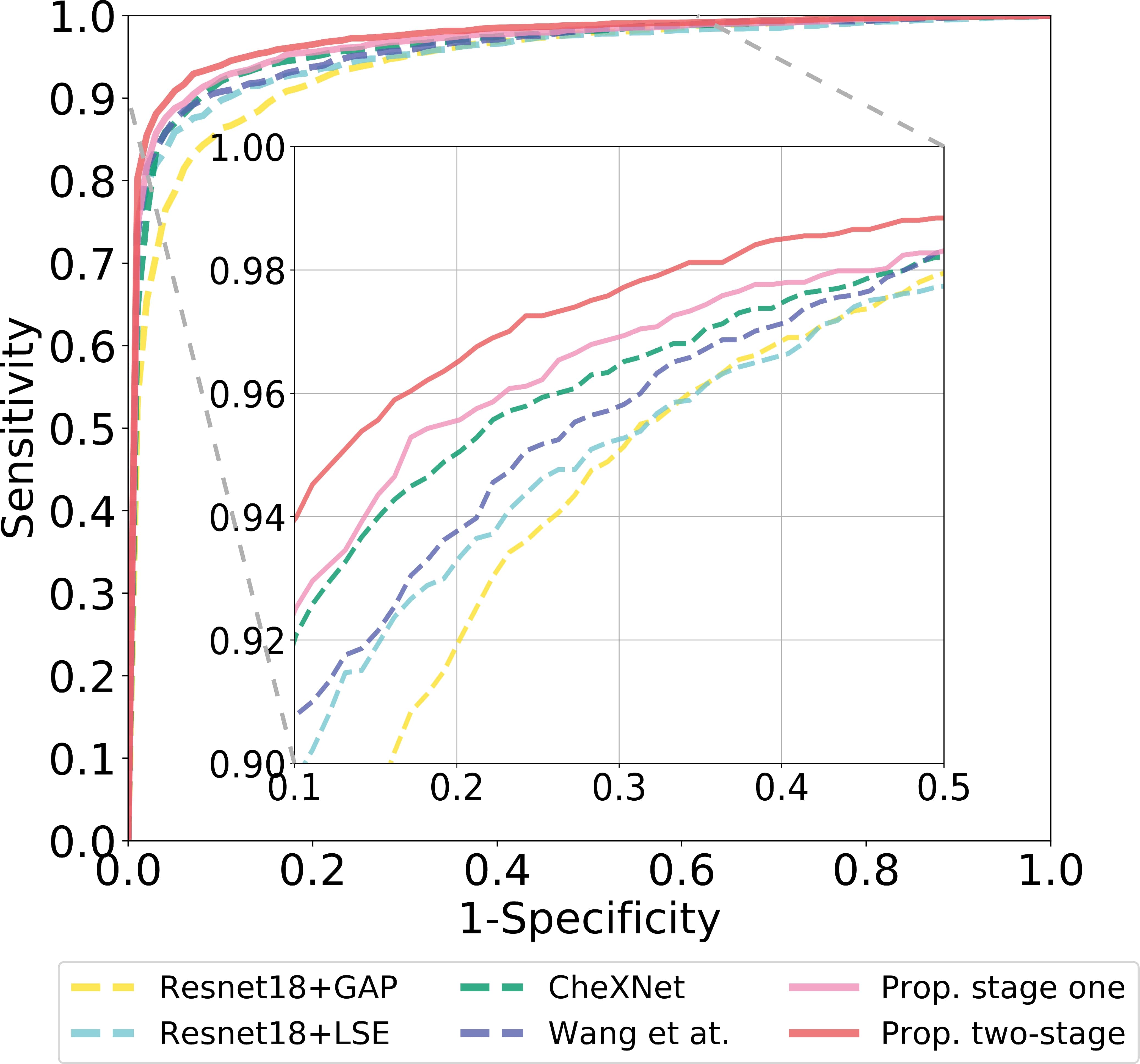
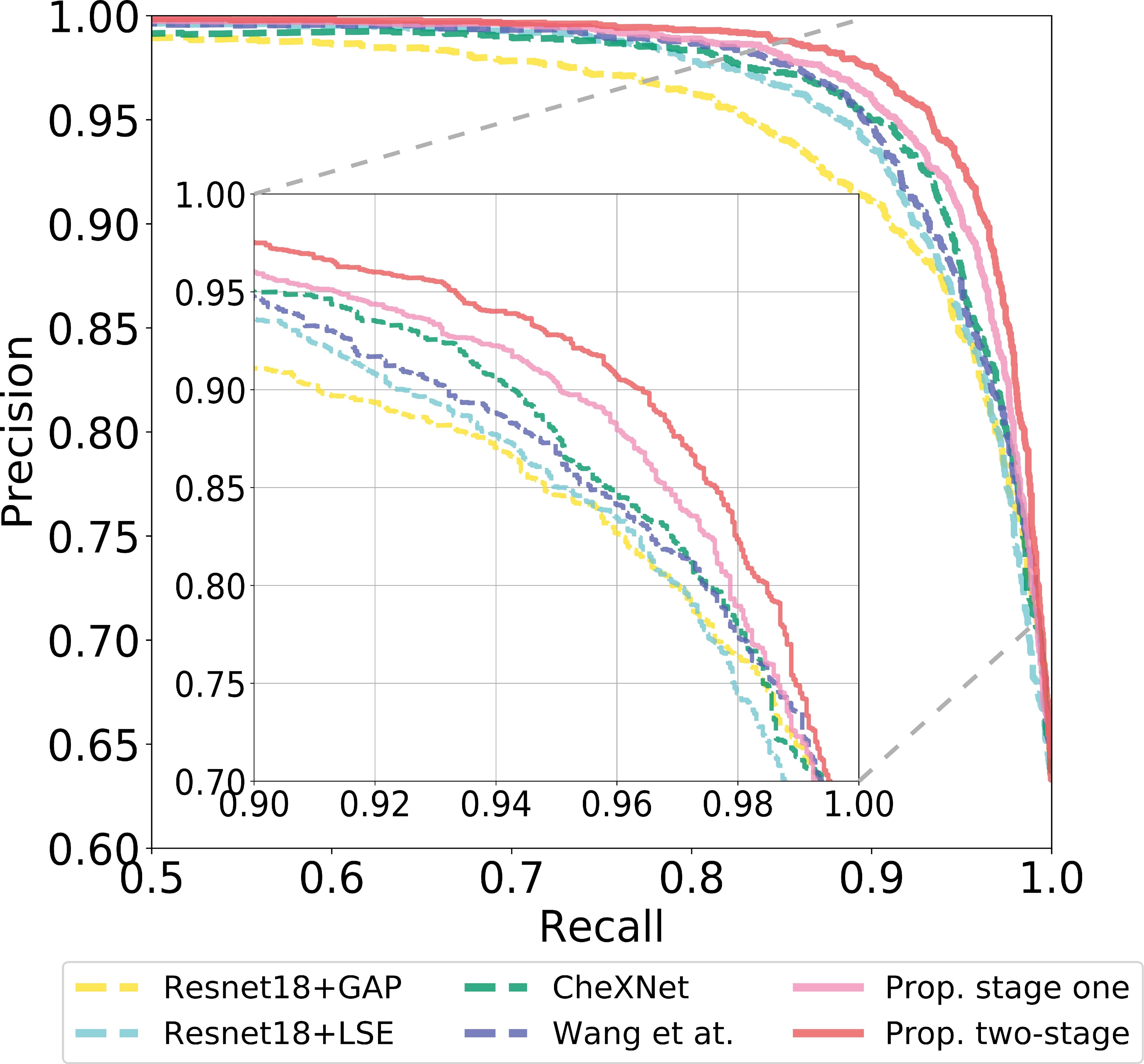
[b] Method AUC \acSR \acRS \acPR ResNet18-GAP 0.946 0.706 0.786 0.846 ResNet18-LSE 0.956 0.723 0.859 0.851 CheXNet [9] 0.962 0.809 0.870 0.876 Wang et al. [11] 0.962 0.752 0.875 0.867 Prop. single-stage 0.968 0.825 0.888 0.903 Prop. two-stage 0.975 0.876 0.909 0.928
Fig. 3 and Tbl. 1 quantitatively summarizes these experiments. As can be seen, all lower-capacity models fare relatively poorly, demonstrating the need to use more descriptive models for global \acPXR interpretation. On the other hand, the first stage of our proposed method achieves an \acAUC of , compared to the achieved by the state-of-the-art single-stage methods [9, 11], demonstrating that our single-stage approach using deep \acMIL can already outperform prior art. With the second stage, our method was able to further improve the \acAUC to , the highest among all evaluated methods. This corresponds to improvements of (), (), and () in \acSR (\acRS) over Wang et al. [11], CheXNet [9], and our single-stage model respectively. These are highly impactful boosts in performance, demonstrating that under high demands of recall and specificity our chained approach can provide drastic improvements.
At our two-stage method achieves a \acPR of , measuring , , and improvements over the two low-capacity baseline models, ResNet18-GAP and ResNet18-LSE, and two high-capacity baseline models CheXNet [9] and Wang et al. [11], respectively. Please note that the actual prevalence of fractures in clinical environments can be lower than our data, which will result in lower precisions for all methods. Nonetheless, the performance ranking would likely to remain, and the improvements of our method over the baselines are expected to be even more significant with a lower prevalence.
In addition, we evaluate the label accuracy of mined probable positive \acpROI on \acpPXR with fracture location annotations, and report a high accuracy of , demonstrating the effectiveness of the proposed weakly-supervised \acROI mining scheme. We also evaluate the hip/pelvic fracture classification performance, and report a high average accuracy of over the five-fold cross validation.
3.2 Reader Study
We conduct reader study to compare performance on \acpPXR with human physicians recruited from the surgical (), orthopedics (), \acER () and radiology () departments. For every \acPXR, physicians were asked to choose from three options: hip fracture, pelvic fracture or no finding. To provide a fair comparison, we used the add-on fracture-type classification output node described in §2.2, which can differentiate between hip and pelvic fractures, matching the three-class classification performed by the readers.
| Accuracy | Hip Fracture | Pelvic Fracture | |||
| Sensitivity | Specificity | Sensitivity | Specificity | ||
| \acsER physician | 0.881 | 0.983 | 0.937 | 0.813 | 0.955 |
| Surgeon | 0.855 | 0.931 | 0.928 | 0.829 | 0.932 |
| Orthopedics specialist | 0.932 | 1.000 | 0.953 | 0.905 | 0.990 |
| Radiologist | 0.930 | 0.990 | 0.965 | 0.870 | 0.995 |
| Physician average | 0.882 | 0.962 | 0.938 | 0.842 | 0.953 |
| Our method | 0.907 | 0.960 | 0.980 | 0.840 | 0.960 |
Tbl. 2 quantitatively summarizes the reader study results. As shown in the results, our method performs comparably to the average physician performance on this dataset, reporting an accuracy of compared to , with higher levels of specificity. Examining the physician specialities in isolation, our method outperforms \acER physicians and surgeons, while not performing as well as the orthopedic specialists and radiologists. Of note, is that when trauma patients are sent to the \acER, it is common that only \acER physicians or surgeons are available to make immediate diagnostic and treatment decisions. As such, the reader study suggests that our approach may be an effective aid for \acPXR fracture diagnosis in the high-stress \acER environment.
4 Conclusion
We introduced a chained two-stage method for universal fracture detection in \acpPXR, consisting of a weakly supervised fracture \acROI mining stage and a localized fracture \acROI classification stage. Experiments show that our method can significantly outperform prior works via five-fold cross validation on \acpPXR. Moreover, a preliminary reader study on \acpPXR involving physicians suggests that our method can perform equivalently to human physicians. Thus, our approach represents an important step forward in automated pelvic and hip fracture diagnosis for \acER environments.
References
- [1] Badgeley, M.A., Zech, J.R., Oakden-Rayner, L., et al.: Deep learning predicts hip fracture using confounding patient and healthcare variables. arXiv:1811.03695 (2018)
- [2] Chellam, W.: Missed subtle fractures on the trauma-meeting digital projector. Injury 47(3), 674–676 (2016)
- [3] Chen, H., Miao, S., Xu, D., Hager, G.D., Harrison, A.P.: Deep hierarchical multi-label classification of chest x-ray images. MIDL (2019)
- [4] Gale, W., Oakden-Rayner, L., Carneiro, G., Bradley, A.P., Palmer, L.J.: Detecting hip fractures with radiologist-level performance using deep neural networks. arXiv:1711.06504 (2017)
- [5] He, K., Zhang, X., Ren, S., Sun, J.: Deep residual learning for image recognition. In: Proceedings of the IEEE conference on computer vision and pattern recognition. pp. 770–778 (2016)
- [6] Huang, G., Liu, Z., Van Der Maaten, L., Weinberger, K.Q.: Densely connected convolutional networks. In: IEEE CVPR. pp. 4700–4708 (2017)
- [7] Jiménez-Sánchez, A., Kazi, A., Albarqouni, S., Kirchhoff, S., Sträter, A., Biberthaler, P., Mateus, D., Navab, N.: Weakly-supervised localization and classification of proximal femur fractures. arXiv:1809.10692 (2018)
- [8] Johnell, O., Kanis, J.: An estimate of the worldwide prevalence, mortality and disability associated with hip fracture. Osteoporosis International 15(11), 897–902 (2004)
- [9] Rajpurkar, P., Irvin, J., Zhu, K., Yang, B., Mehta, H., Duan, T., Ding, D., Bagul, A., Langlotz, C., Shpanskaya, K., et al.: Chexnet: Radiologist-level pneumonia detection on chest x-rays with deep learning. arXiv preprint arXiv:1711.05225 (2017)
- [10] Tarrant, S., Hardy, B., Byth, P., Brown, T., Attia, J., Balogh, Z.: Preventable mortality in geriatric hip fracture inpatients. The bone & joint journal 96(9), 1178–1184 (2014)
- [11] Wang, X., Peng, Y., Lu, L., Lu, Z., Bagheri, M., Summers, R.M.: Chestx-ray8: Hospital-scale chest x-ray database and benchmarks on weakly-supervised classification and localization of common thorax diseases. In: IEEE CVPR (2017)
- [12] Yao, L., Prosky, J., Poblenz, E., Covington, B., Lyman, K.: Weakly supervised medical diagnosis and localization from multiple resolutions. arXiv preprint arXiv:1803.07703 (2018)
- [13] Yuille, A.L., Liu, C.: Deep nets: What have they ever done for vision? CoRR abs/1805.04025 (2018)