Protein recruitment through indirect mechanochemical interactions
Abstract
Some of the key proteins essential for important cellular processes are capable of recruiting other proteins from the cytosol to phospholipid membranes. The physical basis for this cooperativity of binding is, surprisingly, still unclear. Here, we suggest a general feedback mechanism that explains cooperativity through mechanochemical coupling mediated by the mechanical properties of phospholipid membranes. Our theory predicts that protein recruitment, and therefore also protein pattern formation, involves membrane deformation, and is strongly affected by membrane composition.
Protein pattern formation is essential for the spatial organization of intracellular processes Halatek et al. (2018). Examples of biological significance include Min oscillations that guide the positioning of the Z-ring to midcell in E. coli Lutkenhaus (2007), the roles of cell polarization in determining the position of a new growth zone or bud site in S. cerevisiae Johnson (1999) and the anteroposterior axis of the embryo in C. elegans Goldstein and Macara (2007), and spatiotemporal patterns formed by members of the Rho family of GTPases in eukaryotic cells Lawson and Ridley (2018). Such self-organized patterns are the product of a dynamic interplay between diffusion (both in the cytosol and on the membrane) and biochemical reactions among proteins and between proteins and the membrane. A crucial motif in all of the biochemical reaction networks that drive these processes is a nonlinear feedback mechanism, which is generally termed recruitment. Here, membrane-bound proteins facilitate the binding of other soluble proteins from the cytosol to the membrane Halatek et al. (2018). For example, in E. coli, membrane-bound MinD is said to recruit both cytosolic MinD and MinE to the membrane. What then is the physical basis for such cooperative binding between proteins and the membrane? One could adopt a purely chemical perspective and suggest an explanation based on classical concepts of binding cooperativity Hill (1913); Stefan and Le Novère (2013). However, an indiscriminately high chemical affinity between recruiting proteins would also promote protein aggregation in the cytosol as an unwanted side-effect. Then, to still facilitate specific recruitment to the membrane, a possible strategy is for individual proteins to change their conformation upon binding to the membrane so as to become chemically affine scaffolds for other proteins Fischer-Friedrich and Gov (2011); Encinar et al. (2013). In addition to these chemical interactions, binding of proteins to membranes inevitably invokes forces that can lead to membrane deformation.
Here we show how such mechanochemical coupling can lead to a mechanism for the cooperative recruitment of proteins to phospholipid membranes, and thereby provide an alternative strategy for cooperative membrane binding. The basic idea is very simple: Attractive forces between proteins and phospholipids facilitate protein attachment to the membrane. As equal and opposite forces must act on the membrane as well, protein binding will induce mechanical deformation of the membrane. Indeed, it is well known that membrane shape changes can be caused by curvature-inducing polymers and proteins Ford et al. (2002); Tsafrir et al. (2003); Lee et al. (2005); Gov and Gopinathan (2006); Zimmerberg and Kozlov (2006); Prinz and Hinshaw (2009); Stachowiak et al. (2012); McMahon and Boucrot (2015); Jarsch et al. (2016); Gov (2018) containing BAR-domains Zimmerberg and McLaughlin (2004); Peter et al. (2004); Bhatia et al. (2009); Mim and Unger (2012); Zhu et al. (2012); Prévost et al. (2015); Simunovic et al. (2015) and – as recently shown Litschel et al. (2018) – also by the Min family of proteins. Equilibrium theories of the coupling between proteins and membrane generally lead to membrane-mediated interactions between membrane-bound proteins, as reviewed in Phillips et al. (2009); Weikl (2018); Idema and Kraft (2019). The physical origin of such interactions may be hydrophobic mismatch for integral proteins Huang (1986); Wiggins and Phillips (2005); Andersen and Koeppe (2007); Milovanovic et al. (2015); Grau-Campistany et al. (2015), surface interactions that depend on curvature Turner and Sens (2004); Wiggins and Phillips (2005); Iglič et al. (2007); Shlomovitz and Gov (2009); Šárka Perutková et al. (2010); Zhu et al. (2012); Prévost et al. (2015); Mesarec et al. (2016); Agudo-Canalejo and Lipowsky (2017), or membrane shape fluctuations Goulian et al. (1993); Golestanian et al. (1996). Furthermore, these interactions may also depend on the packing density Schäfer et al. (2011) and composition Renner and Weibel (2012); Corradi et al. (2018) of the membrane. Then, proteins that are bound to the membrane effectively attract or repel each other Haselwandter and Phillips (2013); Schweitzer and Kozlov (2015); van der Wel et al. (2016); Vahid and Idema (2016), and form different aggregates Schmidt et al. (2008); Shlomovitz and Gov (2009); Haselwandter and Wingreen (2014); Mesarec et al. (2016); Vahid et al. (2017); Vahid and Idema (2018); Idema and Kraft (2019). Here, however, we do not focus on such self-organization effects. Instead, we ask a different and independent question, namely how membrane deformations affect the affinity and kinetic (un)binding rates of proteins. We propose a general protein recruitment mechanism caused by indirect interactions facilitated through mechanical deformations of the membrane.
As we are interested in quantifying the effect of membrane-mediated interactions on the kinetic rates of protein membrane binding and unbinding, we need to analyze the dynamics of proteins that are subject to both cytosolic diffusion (with diffusion constant ) and a chemical potential gradient caused by the mechanochemical interaction of proteins with the membrane. This is described by a Smoluchowski equation Gardiner (2009); Zwanzig (2001) for the cytosolic protein density :
| (1) |
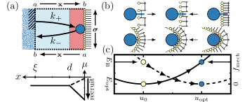
As proteins diffuse freely in the cytosol and interact with the membrane only within some narrow range , a typical spatial profile of the chemical potential is initially flat in the cytosol () and then monotonically approaches that of the proteins at the membrane, , where denotes the free energy functional describing the mechanochemical interaction between proteins and membrane 111 In general, note that this implies that the chemical potential is a function of cytosolic position , and a functional of membrane protein density, . . In general, will depend on both the membrane’s protein density, , and its mechanical state, , at position on the membrane surface; see Fig. 1 for an illustration.
The local free energy density describing the mechanochemical coupling between proteins and the membrane is determined by lipid-lipid and protein-lipid interactions. We assume that a fluid phospholipid membrane can, on a coarse-grained level, be considered as an elastically deformable thin sheet, with bulk modulus , vanishing shear modulus, and a bending modulus, , that is equal for both principal curvatures Helfrich (1973). For low levels of strain, we separate the mechanical degrees of freedom of the membrane into lateral stretching and out-of-plane bending Seifert (1997), and write each mechanical contribution to the free energy as
| (2) |
Here, is a placeholder variable for the mechanical state (conformation) of the membrane, denotes the respective membrane bulk and bending modulus, and denotes the equilibrium conformation (equilibrium density or intrinsic spontaneous curvature 222We further relate our approach to Helfrich’s formulation of the bending energy cost Helfrich (1973) in the SM sup .).
As outlined above, there are several factors that determine the interaction between protein and membrane. Conceptually, one may distinguish between two limiting cases [Fig. 1b]: (A) Protein anchorage through a membrane-targeting domain that penetrates into the inner leaflet of the phospholipid bilayer and induces lateral membrane strain, or (B) protein attachment to the membrane by surface interactions and membrane bending. In both cases, the binding energy, , of a protein to the membrane will depend on the mechanical state (conformation) of the membrane, . In particular, the binding will be strongest, , for some optimal mechanical state, , where it attains an optimal value [Fig. 1c]. This optimal conformation can be understood as a compromise between maximal attractive interactions between proteins and lipids, and minimal steric repulsion [Fig. 1b]. As the membrane becomes crowded with proteins, the binding energy will be reduced due to protein-protein interactions 333Note that the entropic effects of a large protein density can also reduce the protein binding energy, as discussed in the SM sup . There, we show that the general result of nonlinear protein recruitment to the membrane remains valid.. Given that the repulsive part of the Lennard-Jones potential scales as at small distances , this may be accounted for by a factor with ; note that the membrane protein density scales as . Then, a Taylor expansion of the chemical free energy density to lowest order in the membrane conformation, , yields
| (3) |
where the parameter characterizes how strongly the membrane conformation affects protein binding. As noted above, there is a broad range of cytosolic proteins that bind to lipid membranes in a curvature-dependent manner Zimmerberg and McLaughlin (2004); Peter et al. (2004); Bhatia et al. (2009); Mim and Unger (2012); Simunovic et al. (2015); McMahon and Boucrot (2015); Zeno et al. (2018); cf. Fig. 1b, lower panel. For example, protein-curvature coupling can arise from bending proteins to the local membrane curvature Kralj-Iglič et al. (1999); Iglič et al. (2007); Šárka Perutková et al. (2010); Shlomovitz et al. (2011); Bovžič et al. (2015); Prévost et al. (2015); Mesarec et al. (2016), or by bending the membrane to the shape of the proteins in order to maximize attractive interactions [Fig. 1b]. In the following, we specifically consider proteins that couple to the membrane curvature (sum of the two principal curvatures), , and discuss lipid-density-coupling proteins in the SM sup .
As mechanical degrees of freedom relax much faster than protein densities, we adiabatically eliminate the mechanical degrees of freedom by assuming , where 444A further generalization yielding normal and tangential stresses involves variational surface calculus and is briefly outlined in the SM sup . There, we show that the analysis presented here is valid in the limit of small deformations.. This yields a relation between the membrane conformation and the protein density on the membrane: . Here, the ratio between the mechanical modulus , and the mechanochemical coupling parameter , defines a characteristic membrane protein density: . For low membrane protein density, , the interaction between the lipids dominates, and the mechanical state of the membrane is given by the equilibrium value ; cf. yellow symbols in Fig. 1c. With increasing number of attached proteins, the membrane gradually deforms and adopts the mechanical state that is preferred by the proteins; cf. blue symbols in Fig. 1c. There is an interplay between a mechanical energy cost that is lowest at the relaxed state of the membrane, , and a binding energy gain that is highest in the deformed state of the membrane which is optimal for protein binding, . The difference of mechanical free energy density and binding energy between the membrane conformations preferred by the proteins and the lipids read and , respectively.
Upon eliminating the mechanical degrees of freedom using , the interplay between chemical and mechanical terms becomes obvious in the dependence of the free energy density on membrane protein density [Fig. 2a],
| (4) |
where and . The first term encodes free energy costs for membrane deformation through protein binding. With increasing protein density, , this contribution saturates, as the membrane deforms towards a binding-favorable conformation, implying that the corresponding mechanical free energy costs for binding of additional proteins diminish. For intermediate membrane protein densities, the benefit from protein binding (second term in Eq. (4)) dominates. Finally, for very high protein densities, protein binding becomes unfavorable due to crowding ().
The chemical potential at the membrane, , i.e. the energy needed to bind one additional protein to the membrane, reads
| (5) |
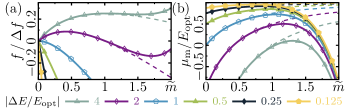
In the absence of crowding effects, the chemical potential approaches the optimal value for large protein densities on the membrane, , meaning that there is an energy gain upon binding [Fig. 2b, dashed lines]. Crowding counteracts this gain, such that protein binding at high densities becomes unfavorable [Fig. 2b, solid lines]. For low densities (), protein binding is also disfavored, as there is a free energy cost for mechanically deforming the membrane that is largest for low membrane protein densities , cf. the last term in Eq. (5). The amplitude of this reduction is given by , which we term the protein binding specificity, as proteins with a higher specificity have a greater preference for mechanical states other than the relaxed state of the membrane [Fig. 2b]. The less specific the binding of a protein, the smaller the changes in the chemical potential as a function of the protein density on the membrane.
What then are the implications of these thermodynamic considerations for the kinetics of protein binding and detachment? To answer this question one has to solve a first-passage-time problem for a particle diffusing in a chemical potential as described by the Smoluchowski equation Eq. (1). This is a well-studied problem, which dates back to Kramers’ theory of reaction kinetics Kramers (1940). For a one-dimensional reaction coordinate , with a reflective boundary at and an absorbing boundary at , the first-passage time is given by Gardiner (2009); Zwanzig (2001):
| (6) |
where is the spatial profile of the chemical potential. In Kramers’ classical escape problem, the reaction rate depends on the height of the barrier that the particle has to cross by diffusion to reach its target Kramers (1940). In our case, however, there is no such barrier. Instead, as discussed above, we expect the landscape to exhibit a monotonically increasing or decreasing profile, depending on whether the chemical potential at the membrane, , is larger or smaller than the value in the bulk of the cytosol (); for an illustration see Fig. 1.
To estimate the kinetic rates, we simplify the geometry of the cell as follows. We divide the space near the membrane into small reaction compartments with respective sizes given by the average distance between proteins, such that each compartment contains a single protein on average. Then, one may approximate a binding process as a one-dimensional diffusion process: an initially unbound protein diffusing in the cytosol enters one of these compartments at a distance from the membrane and after some time encounters the membrane located at . To calculate the corresponding first-passage time, the membrane is considered as an absorbing boundary. The cytosolic boundary of each compartment can effectively be approximated as a reflective boundary, since (on average) there is always one protein within each compartment, i.e. a protein leaving the compartment at is replaced by one entering the compartment. Similarly, an unbinding process may be idealized as a stochastic process, where an initially bound protein detaches at (reflective boundary) and leaves the compartment at (absorbing boundary).
Given our limited knowledge of the profile of the chemical potential, we chose to approximate it by a piecewise linear function [Fig. 1a]. The protein diffuses freely () at large distances from the membrane (). In the vicinity of the membrane (), we assume a linear profile . In the following, we discuss – for simplicity – the case where . The more general (and more realistic) case, where the protein also crosses a preceding flat potential of length , yields qualitatively similar results and is discussed in the SM sup . With these approximations, we can use Eq. (6) to obtain an explicit analytic expression for the mean first-passage times of attachment and detachment Bell and Terentjev (2017). The corresponding kinetic rates, , expressed in units of the basic diffusion time , are found to be
| (7) |
These rates exhibit a pronounced nonlinear dependence on the membrane protein density [Fig. 3]. Hence, protein attachment and detachment are both cooperative processes, owing to the mechanochemical coupling mediated by membrane elasticity.
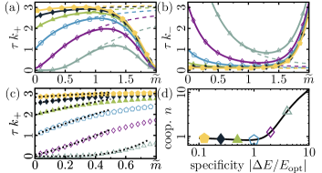
By fitting the attachment rate, Eq. (7), at low densities with , we infer a relationship between the protein specificity and the (Hill) cooperativity coefficient [Fig. 3c, d]; for an analysis in terms of Hill curves please refer to the SM sup . Strong cooperativity () occurs only for high protein specificities, . This implies that induction of a membrane conformation that favors protein binding requires the binding of a disproportionally large number of proteins to the membrane. Therefore, in the deterministic limit, proteins would not attach to the membrane at all [Fig. 2a, empty triangles and diamonds]. However, stochastic binding events, while unlikely at low protein densities, reduce the free energy cost of subsequent binding events and thereby increase their likelihood. This positive feedback leading to recruitment is a purely stochastic effect, and is related to nucleation during discontinuous phase transitions.
To assess whether the proposed indirect cooperativity mechanism could actually come into play at physiological protein concentrations, we estimated its various parameters from known literature values. For proteins with a membrane sensing domain, typical values for the optimal curvature and binding energy are Peter et al. (2004); Wu et al. (2011) and Ma et al. (2017); we assume vanishing spontaneous curvature (). Across different studies, the bending modulus of a phospholipid bilayer was measured to be in the range of , suggesting a typical value Phillips et al. (2009); Dimova (2014); Nagle et al. (2015). Taking a value for protein specificity where nonlinear binding kinetics is significant (recruitment) [Fig. 3d], the corresponding range of concentrations, , easily encompasses any physiological value; the maximum packing density of proteins with size is .
In summary, we have shown that mechanochemical coupling between proteins provides a possible mechanism for the nonlinear binding kinetics (recruitment) of proteins to the membrane.
The effect originates from the interplay between protein-lipid and lipid-lipid interactions, which induce mechanical deformations of the membrane and thereby alter the protein binding environment.
As protein-lipid interactions become dominant with increasing concentrations of membrane-bound proteins, the membrane’s mechanical state becomes more favorable for binding.
This shows how cooperativity and the recruitment of proteins can naturally emerge without any reliance on direct chemical interactions and conformational changes.
The results should certainly be applicable to proteins that are known to bend membranes, e.g. proteins containing BAR domains Zimmerberg and McLaughlin (2004); Peter et al. (2004); Bhatia et al. (2009); Mim and Unger (2012); Simunovic et al. (2015).
As recent experiments have unexpectedly shown that Min protein oscillations can lead to oscillations in vesicle shape Litschel et al. (2018), we would argue that our theory should also apply to the broad class of NTPases that are essential for cellular protein pattern formation.
Thus, strain sensing and generation might not only be a property of a few specialized proteins, but might actually be a prominent and perhaps general feature of membrane-binding proteins.
Further exploration of curvature sensing during macroscopic pattern formation might be highly rewarding Peleg et al. (2011); Thalmeier et al. (2016); Wu et al. (2018).
Our theory predicts that one can alter the recruitment exponent of membrane-binding proteins by tuning the protein specificity (possibly by changing the membrane composition or introducing permanently-bound membrane-bending proteins).
Such a change in cooperativity should have a much stronger effect on emerging protein patterns than the tuning of reaction rates, because it changes the nature of the nonlinear coupling.
We would expect profound changes in the protein dynamics that could be explored using appropriately modified reaction-diffusion models for various cellular systems Huang et al. (2003); Halatek and Frey (2012); Klünder et al. (2013); Denk et al. (2018); Goryachev and Leda (2017); Halatek and Frey (2018), as well as by experimentally tinkering with the composition of the membrane.
Finally, it would be highly interesting and rewarding to quantify the mechanochemical effect for specific membrane-binding proteins experimentally.
This would provide an interesting basis for theoretical models of pattern-forming protein systems and contribute towards revealing the universal role of membrane elasticity in cellular functions.
Acknowledgements.
We thank Fridtjof Brauns, George Dadunashvili, Raphaela Geßele, Igor Goychuk, Isabella Graf, Laeschkir Hassan, Timon Idema, Anatoly B. Kolomeisky, Thomas Litschel, Rüdiger Thul and Manon Wigbers for stimulating discussions. E.F. acknowledges financial support from the Deutsche Forschungsgemeinschaft (DFG) via the Collaborative Research Center (SFB) 1032 (project B2). A.G. is supported by a DFG fellowship through the Graduate School Quantitative Biosciences Munich (QBM). E.F. also acknowledges the hospitality of the Kavli Institute of Nanoscience at TU Delft, where part of this work was done.References
- Halatek et al. (2018) J. Halatek, F. Brauns, and E. Frey, Philos. Trans. Royal Soc. B 373, 20170107 (2018).
- Lutkenhaus (2007) J. Lutkenhaus, Annu. Rev. Biochem. 76, 539 (2007).
- Johnson (1999) D. I. Johnson, Microbiol. Mol. Biol. Rev. 63, 54 (1999).
- Goldstein and Macara (2007) B. Goldstein and I. G. Macara, Dev. Cell 13, 609 (2007).
- Lawson and Ridley (2018) C. D. Lawson and A. J. Ridley, J. Cell Biol. 217, 447 (2018).
- Hill (1913) A. V. Hill, Biochem. J. 7, 471 (1913).
- Stefan and Le Novère (2013) M. I. Stefan and N. Le Novère, PLOS Comp. Biol. 9, 1 (2013).
- Fischer-Friedrich and Gov (2011) E. Fischer-Friedrich and N. Gov, Phys. Biol. 8, 026007 (2011).
- Encinar et al. (2013) M. Encinar, A. V. Kralicek, A. Martos, M. Krupka, S. Cid, A. Alonso, A. Rico, I., M. Jiménez, and M. Vélez, Langmuir 29, 9436 (2013).
- Ford et al. (2002) M. G. J. Ford, I. G. Mills, B. J. Peter, Y. Vallis, G. J. K. Praefcke, P. R. Evans, and H. T. McMahon, Nature 419, 361 (2002).
- Tsafrir et al. (2003) I. Tsafrir, Y. Caspi, M.-A. Guedeau-Boudeville, T. Arzi, and J. Stavans, Phys. Rev. Lett. 91, 138102 (2003).
- Lee et al. (2005) M. C. Lee, L. Orci, S. Hamamoto, E. Futai, M. Ravazzola, and R. Schekman, Cell 122, 605 (2005).
- Gov and Gopinathan (2006) N. S. Gov and A. Gopinathan, Biophys. J. 90, 454 (2006).
- Zimmerberg and Kozlov (2006) J. Zimmerberg and M. M. Kozlov, Nat. Rev. Mol. Cell Biol. 7, 9 (2006).
- Prinz and Hinshaw (2009) W. A. Prinz and J. E. Hinshaw, Crit. Rev. Biochem. Mol. Biol. 44, 278 (2009).
- Stachowiak et al. (2012) J. C. Stachowiak, E. M. Schmid, C. J. Ryan, H. S. Ann, D. Y. Sasaki, M. B. Sherman, P. L. Geissler, D. A. Fletcher, and C. C. Hayden, Nat. Cell Biol. 14, 944– (2012).
- McMahon and Boucrot (2015) H. T. McMahon and E. Boucrot, J. Cell Sci. 128, 1065 (2015).
- Jarsch et al. (2016) I. K. Jarsch, F. Daste, and J. L. Gallop, J. Cell Biol. 214, 375 (2016).
- Gov (2018) N. S. Gov, Philosophical Transactions of the Royal Society B: Biological Sciences 373, 20170115 (2018).
- Zimmerberg and McLaughlin (2004) J. Zimmerberg and S. McLaughlin, Curr. Biol. 14, R250 (2004).
- Peter et al. (2004) B. J. Peter, H. M. Kent, I. G. Mills, Y. Vallis, P. J. G. Butler, P. R. Evans, and H. T. McMahon, Science 303, 495 (2004).
- Bhatia et al. (2009) V. K. Bhatia, K. L. Madsen, P.-Y. Bolinger, A. Kunding, P. Hedegård, U. Gether, and D. Stamou, EMBO J. 28, 3303 (2009).
- Mim and Unger (2012) C. Mim and V. M. Unger, Trends Biochem. Sci. 37, 526 (2012).
- Zhu et al. (2012) C. Zhu, S. L. Das, and T. Baumgart, Biophys. J. 102, 1837 (2012).
- Prévost et al. (2015) C. Prévost, H. Zhao, J. Manzi, E. Lemichez, P. Lappalainen, A. Callan-Jones, and P. Bassereau, Nat. Commun. 6, 8529 (2015).
- Simunovic et al. (2015) M. Simunovic, G. A. Voth, A. Callan-Jones, and P. Bassereau, Trends Cell Biol. 25, 780 (2015).
- Litschel et al. (2018) T. Litschel, B. Ramm, R. Maas, M. Heymann, and P. Schwille, Angew. Chem. Int. Ed. 57, 16286 (2018).
- Phillips et al. (2009) R. Phillips, T. Ursell, P. Wiggins, and P. Sens, Nature 459, 379 (2009).
- Weikl (2018) T. R. Weikl, Annu. Rev. Phys. Chem. 69, 521 (2018).
- Idema and Kraft (2019) T. Idema and D. J. Kraft, Curr. Opin. Colloid Interface Sci. 40, 58 (2019).
- Huang (1986) H. Huang, Biophys. J. 50, 1061 (1986).
- Wiggins and Phillips (2005) P. Wiggins and R. Phillips, Biophys. J. 88, 880 (2005).
- Andersen and Koeppe (2007) O. S. Andersen and R. E. Koeppe, Annu. Rev. Biophys. Biomol. Struct. 36, 107 (2007).
- Milovanovic et al. (2015) D. Milovanovic, A. Honigmann, S. Koike, F. Göttfert, G. Pähler, M. Junius, S. Müllar, U. Diederichsen, A. Janshoff, H. Grubmüller, H. J. Risselada, C. Eggeling, S. W. Hell, G. van den Bogaart, and R. Jahn, Nature Comm. 6, 5984 (2015).
- Grau-Campistany et al. (2015) A. Grau-Campistany, E. Strandberg, P. Wadhwani, J. Reichert, J. Bürck, F. Rabanal, and A. S. Ulrich, Sci. Rep. 5, 9388 (2015).
- Turner and Sens (2004) M. S. Turner and P. Sens, Phys. Rev. Lett. 93, 118103 (2004).
- Iglič et al. (2007) A. Iglič, T. Slivnik, and V. Kralj-Iglič, J. Biomech. 40, 2492 (2007).
- Shlomovitz and Gov (2009) R. Shlomovitz and N. S. Gov, Phys. Biol. 6, 046017 (2009).
- Šárka Perutková et al. (2010) Šárka Perutková, V. Kralj-Iglič, M. Frank, and A. Iglič, J. Biomech. 43, 1612 (2010).
- Mesarec et al. (2016) L. Mesarec, W. Góźdź, V. K. Iglič, S. Kralj, and A. Iglič, Colloids Surf. B 141, 132 (2016).
- Agudo-Canalejo and Lipowsky (2017) J. Agudo-Canalejo and R. Lipowsky, Soft Matter 13, 2155 (2017).
- Goulian et al. (1993) M. Goulian, R. Bruinsma, and P. Pincus, Europhys. Lett. 22, 145 (1993).
- Golestanian et al. (1996) R. Golestanian, M. Goulian, and M. Kardar, Europhys. Lett. 33, 241 (1996).
- Schäfer et al. (2011) L. V. Schäfer, D. H. de Jong, A. Holt, A. J. Rzepiela, A. H. de Vries, B. Poolman, J. A. Killian, and S. J. Marrink, Proc. Natl. Acad. Sci. U.S.A. 108, 1343 (2011).
- Renner and Weibel (2012) L. D. Renner and D. B. Weibel, J. Biol. Chem. 287, 38835 (2012).
- Corradi et al. (2018) V. Corradi, E. Mendez-Villuendas, H. I. Ingólfsson, R.-X. Gu, I. Siuda, M. N. Melo, A. Moussatova, L. J. DeGagné, B. I. Sejdiu, G. Singh, T. A. Wassenaar, K. Delgado Magnero, S. J. Marrink, and D. P. Tieleman, ACS Cent. Sci. 4, 709 (2018).
- Haselwandter and Phillips (2013) C. A. Haselwandter and R. Phillips, Europhys. Lett. 101, 68002 (2013).
- Schweitzer and Kozlov (2015) Y. Schweitzer and M. M. Kozlov, PLOS Comp. Biol. 11, 1 (2015).
- van der Wel et al. (2016) C. van der Wel, A. Vahid, A. Šarić, T. Idema, D. Heinrich, and D. J. Kraft, Sci. Rep. 6, 32825 (2016).
- Vahid and Idema (2016) A. Vahid and T. Idema, Phys. Rev. Lett. 117, 138102 (2016).
- Schmidt et al. (2008) U. Schmidt, G. Guigas, and M. Weiss, Phys. Rev. Lett. 101, 128104 (2008).
- Haselwandter and Wingreen (2014) C. A. Haselwandter and N. S. Wingreen, PLOS Comp. Biol. 10, 1 (2014).
- Vahid et al. (2017) A. Vahid, A. Šarić, and T. Idema, Soft Matter 13, 4924 (2017).
- Vahid and Idema (2018) A. Vahid and T. Idema, bioRxiv 10.1101/336545 (2018).
- Gardiner (2009) C. Gardiner, Stochastic Methods (Springer Berlin Heidelberg, 2009).
- Zwanzig (2001) R. Zwanzig, Nonequilibrium Statistical Mechanics (Oxford University Press, 2001).
- Note (1) In general, note that this implies that the chemical potential is a function of cytosolic position , and a functional of membrane protein density, .
- Helfrich (1973) W. Helfrich, Z. Naturforsch. C Bio. Sci. 28, 693 (1973).
- Seifert (1997) U. Seifert, Adv. Phys. 46, 13 (1997).
- Note (2) We further relate our approach to Helfrich’s formulation of the bending energy cost Helfrich (1973) in the SM sup .
- Note (3) Note that the entropic effects of a large protein density can also reduce the protein binding energy, as discussed in the SM sup . There, we show that the general result of nonlinear protein recruitment to the membrane remains valid.
- Zeno et al. (2018) W. F. Zeno, U. Baul, W. T. Snead, A. C. M. DeGroot, L. Wang, E. M. Lafer, D. Thirumalai, and J. C. Stachowiak, Nature Comm. 9, 4152 (2018).
- Kralj-Iglič et al. (1999) V. Kralj-Iglič, V. Heinrich, S. Svetina, and B. Žekš, Eur. Phys. J. B 10, 5 (1999).
- Shlomovitz et al. (2011) R. Shlomovitz, N. S. Gov, and A. Roux, New J. Phys. 13, 065008 (2011).
- Bovžič et al. (2015) B. Bovžič, S. L. Das, and S. Svetina, Soft Matter 11, 2479 (2015).
- (66) See Supplemental Material at [URL will be inserted by publisher] for further details and an additional analysis of the model, which includes Refs. Zhong-Can and Helfrich (1989); Doi (2013); Deserno (2015); Guckenberger and Gekle (2017); Goychuk et al. .
- Note (4) A further generalization yielding normal and tangential stresses involves variational surface calculus and is briefly outlined in the SM sup . There, we show that the analysis presented here is valid in the limit of small deformations.
- Kramers (1940) H. A. Kramers, Physica 7, 284 (1940).
- Bell and Terentjev (2017) S. Bell and E. M. Terentjev, Biophys. J. 112, 2439 (2017).
- Wu et al. (2011) W. Wu, K.-T. Park, T. Holyoak, and J. Lutkenhaus, Mol. Microbiol. 79, 1515 (2011).
- Ma et al. (2017) L. Ma, Y. Cai, Y. Li, J. Jiao, Z. Wu, B. O’Shaughnessy, P. De Camilli, E. Karatekin, and Y. Zhang, eLife 6, e30493 (2017).
- Dimova (2014) R. Dimova, Adv. Colloid Interface Sci. 208, 225 (2014).
- Nagle et al. (2015) J. F. Nagle, M. S. Jablin, S. Tristram-Nagle, and K. Akabori, Chem. Phys. Lipids 185, 3 (2015).
- Peleg et al. (2011) B. Peleg, A. Disanza, G. Scita, and N. Gov, PLOS ONE 6, 1 (2011).
- Thalmeier et al. (2016) D. Thalmeier, J. Halatek, and E. Frey, Proc. Natl. Acad. Sci. U.S.A. , 201515191 (2016).
- Wu et al. (2018) Z. Wu, M. Su, C. Tong, M. Wu, and J. Liu, Nat. Commun. 9, 136 (2018).
- Huang et al. (2003) K. C. Huang, Y. Meir, and N. S. Wingreen, Proc. Natl. Acad. Sci. U.S.A. 100, 12724 (2003).
- Halatek and Frey (2012) J. Halatek and E. Frey, Cell Rep. 1, 741 (2012).
- Klünder et al. (2013) B. Klünder, T. Freisinger, R. Wedlich-Söldner, and E. Frey, PLOS Comp. Biol. 9, 1 (2013).
- Denk et al. (2018) J. Denk, S. Kretschmer, J. Halatek, C. Hartl, P. Schwille, and E. Frey, Proc. Natl. Acad. Sci. U.S.A. 115, 4553 (2018).
- Goryachev and Leda (2017) A. B. Goryachev and M. Leda, Mol. Biol. Cell 28, 370 (2017).
- Halatek and Frey (2018) J. Halatek and E. Frey, Nat. Phys. 14, 507 (2018).
- Zhong-Can and Helfrich (1989) O.-Y. Zhong-Can and W. Helfrich, Phys. Rev. A 39, 5280 (1989).
- Doi (2013) M. Doi, Soft Matter Physics (Oxford University Press, 2013).
- Deserno (2015) M. Deserno, Chem. Phys. Lipids 185, 11 (2015).
- Guckenberger and Gekle (2017) A. Guckenberger and S. Gekle, J. Phys. Condens. Matter 29, 203001 (2017).
- (87) A. Goychuk, L. Hassan, M. Wigbers, and E. Frey, in preparation .