Core-level x-ray photoemission and Raman spectroscopy studies on electronic structures in Mott-Hubbard type nickelate oxide NdNiO2
Abstract
We perform core-level X-ray photoemission spectroscopy (XPS) and electronic Raman scattering studies of electronic structures and spin fluctuations in the bulk samples of the nickelate oxide NdNiO2. According to Nd and O XPS spectra, we conclude that NdNiO2 has a large transfer energy. From the analysis of the main line of the Ni XPS, we confirm the NiO2 planes in NdNiO2 are of Mott-Hubbard type in the Zaanen-Sawatzky-Allen scheme. The two-magnon peak in the Raman scattering provides direct evidence for the strong spin-fluctuation in NdNiO2. The peak position determines the antiferromagnetic exchange meV. Our experimental results agree well with our previous theoretical results.
Introduction – Nickel has the formal valences Ni3+, Ni2+ and Ni+ with electronic configurations of of 3 , 3 and 3d9, respectively, in the transition-metal oxides. The nickelate compounds exhibit lots of strongly correlated electronic properties, such as the metal-insulator transitions, unusual magnetic order, charge order and even superconductivity Zaanen et al. (1985); Medarde (1997); Catalan (2008); Middey et al. (2016); Catalano et al. (2018); Crespin et al. (1983); Hayward et al. (1999); Hayward and Rosseinsky (2003); Anisimov et al. (1999); Lee and Pickett (2004); Kawai et al. (2009); Kaneko et al. (2009); Kawai et al. (2010); Ikeda et al. (2013, 2016); Li et al. (2019); Hepting et al. (2020). The recent discovery of the superconductivity in the thin film Nd0.8Sr0.2NiO2 Li et al. (2019) has the similar crystal and 3 electronic structures to the cuprate superconductors Bednorz and Müller (1986), and motivates the experimental Hepting et al. (2020) and lots of theoretical works Botana and Norman (2020); Sakakibara et al. (2019); Jiang et al. (2019a); Wu et al. (2020); Nomura et al. (2019); Gao et al. (2019); Zhang et al. (2020a); Ryee et al. (2020); Zhang et al. (2020b); Zhang and Vishwanath (2019); Jiang et al. (2019b); Hu and Wu (2019); Chang et al. (2019); Werner and Hoshino (2020); Liu et al. (2019); Choi et al. (2020); Karp et al. (2020) to explore its electronic properties. The recent superconductivity dome spanning the doping concentration 0.1250.25 in thin-film Nd1-xSrxNiO2 infers the remarkable similarity to cuprate superconductors Li et al. (2020).
Reduced form of perovskite nickelate LaNiO3 and NdNiO3 leads to the infinite layered phase LaNiO2 and NdNiO2 Crespin et al. (1983); Hayward et al. (1999); Hayward and Rosseinsky (2003); Kawai et al. (2009); Kaneko et al. (2009); Ikeda et al. (2013, 2016). Several experimental studies have been carried out on the parent superconducting nickelate oxides. Magnetization measurements and powder neutron diffraction were performed on LaNiO2 and NdNiO2 to study the magnetic properties Hayward et al. (1999); Hayward and Rosseinsky (2003). Electric transport measurements have been performed on the thin film samples LaNiO2 and NdNiO2, and the resistivity exhibits the insulator or semiconductor behavior depending on the samples Kawai et al. (2009); Kaneko et al. (2009); Ikeda et al. (2013, 2016); Li et al. (2019). The Ni-K edge X-ray absorption spectroscopy (XAS) was implemented to characterize the LaNiO2 samples Crespin et al. (1983); Kawai et al. (2009). Recently, with the help of density functional theory within LDA+U scheme, Hepting et. al. Hepting et al. (2020) has used XAS and X-ray emission spectroscopy (XES) to unveil a large charge-transfer energy, and categorized LaNiO2 and NdNiO2 to the Mott-Hubbard type according to the Zaanen-Sawatzky-Allen (ZSA) scheme Zaanen et al. (1985). The Mott-Hubbard scenario is still under debate, and the charge-transfer type is also proposed for NdNiO2 Karp et al. (2020).
In this paper, the main purpose to study electronic structures in NdNiO2 is twofold. Firstly, we perform the core-level x-ray photoemission spectroscopy (XPS) to examine the existence of the Zhang-Rice singlet Zhang and Rice (1988) to address the ZSA classification issue for NdNiO2. Ni+ has higher chemical potential and hence a larger transfer energy in NdNiO2 as observed in XAS and XES Hepting et al. (2020). We implement the XPS measurement to further disprove the large- charge-transfer (a charge-transfer insulator with a large transfer energy ) scenario for NdNiO2. The main line of the transition-metal XPS spectrum has the doubly peaked structure in the cuprate parent compound Fujimori et al. (1987); Shen et al. (1987) and NiO van Veenendaal and Sawatzky (1993) due to the non-local screening effects van Veenendaal and Sawatzky (1993); van Veenendaal (2006); Ghiasi et al. (2019), related to the Zhang-Rice singlet in charge-transfer insulators Zhang and Rice (1988). In the XPS measurements, from Nd 3 and O 1 XPS, we find NdNiO2 has a larger transfer energy , comparing to a small/negative charge-transfer insulator NdNiO3. The non-local screening doubly peaked structure of the main line of Ni 2p3/2 is significantly suppressed in NdNiO2, confirming the Mott-Hubbard scenario for NdNiO2 Hepting et al. (2020).
Secondly, as the NiO2 planes in NdNiO2 is in the Mott-Hubbard regime in the ZSA scheme, we are concerned with the spin dynamics in the system. The strong antiferromagnetic spin fluctuation is generally accepted as a key ingredient for the unconventional superconductivity in the cuprate oxides, due to the proximity to a Mott insulator Anderson (1987); Lee et al. (2006). Raman spectroscopy probes spin fluctuations in the transition-metal compounds, even without a long-range magnetic order Devereaux and Hackl (2007). The exchange interaction for cuprates is very large, and was determined from the two-magnon peak to be of order 125 meV Sugai et al. (1988); Lyons et al. (1988, 1989); Knoll et al. (1990); Sulewski et al. (1990, 1991); Blumberg et al. (1994, 1996); Rübhausen et al. (1997, 1999); Sugai et al. (2003); Devereaux and Hackl (2007). The absence of long-range magnetic order in LaNiO2 and NdNiO2 in Refs. Hayward et al. (1999) may be due to poor sample qualities, or due to the “self-doping” effects of the Nd electron pockets Lee and Pickett (2004); Hepting et al. (2020); Zhang et al. (2020a). We perform Raman measurements to explore spin fluctuations, and observe a broad two-magnon peak in NdNiO2. The antiferromagnetic exchange is determined as meV in NdNiO2, close to our previous theoretical estimation Zhang et al. (2020a).
Experimental setup. – We carried out the XPS investigations on Thermo Fisher ESCALAB 250Xi using monochromated Al K radiation at room temperature, and the electron flood gun was turned on to eliminate electric charging effect in our insulating samples. The binding energy in XPS was calibrated by spectra of carbon. The Shirley background of the XPS spectra has been subtracted in this work. The energy dispersive X-ray (EDX) and scanning electron microscopy (SEM) studies were performed on Phenom ProX equipped with Backscatter detector at an operating voltage of 15 kV. X-ray diffraction (XRD) measurements were conducted on Rigaku Smartlab 9 KW using Cu K radiation at room temperature. The Raman spectra were measured in the quasi-back-scattering geometry on our home-built system using a HORIBA iHR550 spectrometer and the 632.8 nm excitation line of a He-Ne laser. The power of the laser is about 400 W, and we set 1200 grooves/mm grating and 30 min integral time. The samples were placed in a He-flow cryostat which evacuated to Torr.
Single crystal synthesis. –
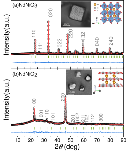
The NdNiO3 polycrystals were synthesized by a combination precursor method with high pressure technique. The precursor was prepared by sintering the sol-gel with stoichiometric ratio of Ni and Nd Vassiliou et al. (1989). After calcining the precursor powder capsuled in h-BN at 1073 K under high pressure of 2 GPa, the crystals of NdNiO3 with high quality were successfully obtained, as shown in Fig. 1. The reduction of NdNiO3 was performed at low temperature using CaH2 as the reducing agent, as reported in refs Kawai et al. (2009); Li et al. (2019), and finally, the phase of NdNiO2 was obtained.
We observe the broadening of the peak width in the XRD pattern for NdNiO2, especially in the peak indexed by with nonzero value for . The anisotropic broadening of the Bragg peaks was also observed in both thin films and polycrystals Hayward et al. (1999); Hayward and Rosseinsky (2003); Kawai et al. (2009); Kaneko et al. (2009); Li et al. (2019, 2019), and could be ascribed to the anisotropic crystal size or stacking disorder. NdNiO2 has intensive peaks along direction in XRD pattern as shown in Fig. 1 (b), probably due to the preferred -axis of crystal grains. The orientation preference was reported for the thin-film LaNiO2 where the preferred orientation changes from -axis to -axis for the over-time reduction Kaneko et al. (2009); Kawai et al. (2010).
We performed the Rietveld profile refinement for NdNiO3 and NdNiO2 using the Fullprof suite of programs Rodríguez-Carvajal (1993). The NdNiO3 has the space group of with lattice parameters Å, Å, and Å (% and %), consistent with the previous report García-Muñoz et al. (2009). The refinement results for NdNiO2 is Å and Å ( with % and % ), which is in agreement with previous results which used NaH as reduction agent Hayward and Rosseinsky (2003). It is worth noting that the metal nickel phase usually appears in NdNiO2 after reduction, as reported in refs Hayward et al. (1999); Hayward and Rosseinsky (2003); Kawai et al. (2009); Li et al. (2019). Although the metal nickel phase is almost invisible in our PXRD result, we observed the metal nickel impurities embedded in surface-polished NdNiO2 crystal by SEM, as shown in inset of Fig. 5.
Electronic structures from XPS. – The core-level x-ray spectroscopy is a powerful tool to study electronic properties of transition-metal oxides De Groot and Kotani (2008); Van Veenendaal (2015). It helps to determine the on-site Coulomb interaction and the charge-transfer energy, and classify materials as charge-transfer or Mott-Hubbard insulators in the Zaanen-Sawatzky-Allen scheme van der Laan et al. (1981); Gunnarsson and Schönhammer (1983); Zaanen et al. (1985, 1986); Bocquet et al. (1992).
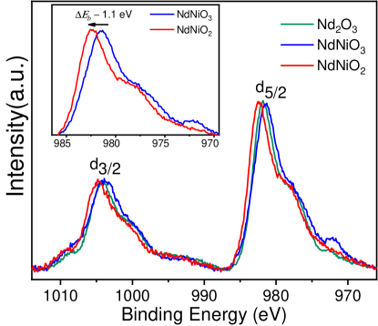
Figure 2 is the Nd 3 core-level XPS spectra for Nd2O3, NdNiO3 and NdNiO2. The similar overall pattern indicates the trivalent Nd3+ with the 4 ground state in the three compounds. There is a blue shift of the binding energy around eV for NdNiO2, comparing to NdNiO3 as shown in the inset. Thus the formal chemical valence is Ni3+ and Ni1+ in NdNiO3 and NdNiO2, respectively. In the Nd3+ XPS, the spectral structure is mainly determined by the effect of the covalency hybridization, and the two peaks of the XPS correspond to the bonding and antibonding states between the and configurations (( is the hole in the ligand) in the final states De Groot and Kotani (2008). The ground state of Nd2O3 is in the almost pure 4 configuration, and hence the main peak at around 981-983 eV in the Nd 3 XPS spectra is the unscreened final state. The screened final state has a lower binding energy around 977 eV.
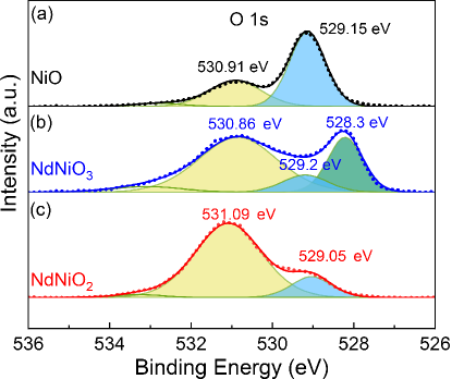
Figure 3 compares the O 1 XPS spectra in NiO, NdNiO3 and NdNiO2. The O 1 XPS has often used in the experiments for the transition-metal oxides, however, the nature of the 531 eV O 1 spectral peak in NiO is unclear Sarma and Chainani (1990); Biesinger et al. (2009), and has been proposed to be due to defective sites within the oxide crystal Hagelin-Weaver et al. (2004); Payne et al. (2009), adsorbed oxygen Benndorf et al. (1982), or hydroxide species Carley et al. (1983), and also attributed to an intrinsic feature Weaver et al. (1988); Sarma and Chainani (1990). We can’t exclude the contaminant origin for the 530.9 eV feature, however, we have measure the O 1 XPS spectra on both different samples and different XPS instruments and obtain the consistent similar results, which is hardly attributed to an extrinsic feature. In O XPS spectra, NiO and NdNiO2 have the best-screened states at around 529 eV. In NdNiO3, an extra peak appears at the lowest binding energy 528.3 eV, indicating the concentration of the oxygen holes in the ground state, due to a negative charge transfer energy Johnston et al. (2014); Mizokawa et al. (1991, 1995); Abbate et al. (2002).
As depicted in the inset of Fig. 2, NdNiO2 has a higher binding energy than NdNiO3 in the Nd XPS by around eV. The Nd3+ binding energy blue shift indicates the increasing of the Fermi energy in NdNiO2. Since Nd3+ has a weaker hybridization than Ni+ with the oxygen, the Fermi energy increasing is mainly due to the increasing chemical potential of Ni+ in NdNiO2, assuming the unchanged chemical potential of oxygen. Hence NdNiO2 has a larger charge-transfer energy . Taking account for the covalency of the transition-metal ions and ligands, the effective charge transfer energy is Bisogni et al. (2016), where and are the bandwidths for the transition-metal 3-band and oxygen 2-band, respectively. NdNiO2 has the nearly two-dimensional transition-metal 3-band and oxygen 2-band which are narrower than the three-dimensional ones in NdNiO3; then it has has a larger effective charge transfer energy . From the comparison between NdNiO2 and NdNiO3, the former has a large charge-transfer energy than the latter, and is not a negative/small charge-transfer insulator. NdNiO2 has the quite similar pattern of O 1 XPS to NiO, but the covalence peak is significantly suppressed as shown in Fig. 3. Since NiO is a three-dimensional charge-transfer insulator, NdNiO2 could have a larger effective charge-transfer energy than NiO, and is likely to be a Mott-Hubbard type as suggested in Ref. Li et al. (2019); Hepting et al. (2020); Jiang et al. (2019a); Zhang et al. (2020a), which need further confirmation in the Ni XPS as demonstrated below.
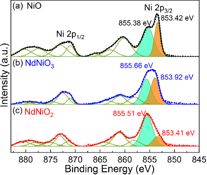
Next, we examine the Ni 2 XPS spectra to confirm whether NdNiO2 is a charge-transfer or Mott-Hubbard type. Figure 4 displays Ni 2 XPS spectra of NiO, NdNiO3 and NdNiO2. The Ni 2p spectrum is composed of 2 and 2 components due to the large spin-orbit coupling in the Ni core level. The main line region of the spectra around 852-859 eV corresponds to the screened final states, and can be fitted by two peaks. The removal of the 2 core electron leads to the creation of a strong local potential on the nickel site, and an electron is transferred from the environment to the Ni site. The different types, i.e., small/negative charge-transfer, charge-transfer and Mott-Hubbard types according to the ZSA scheme, of the transition-metal compounds have different non-local screening effects in the main line of the transition-metal XPS spectra van Veenendaal and Sawatzky (1993); van Veenendaal (2006).
The perovskite NdNiO3 has a negative effective charge-transfer energy, giving rise to “self-doped” holes on the ligands Bisogni et al. (2016). It mainly has a local Ni 3 configuration, a predominant O 2 character across the Fermi level and a consequent ground state of mainly Ni 3 in the metallic state at the room temperature. The two peaks in the main lines correspond to the and final states Mizokawa et al. (1995). NdNiO2 has a larger transfer energy than NdNiO3 as demonstrated above according to Fig. 2 and Fig. 3, and hence displays a different shape of the main line of Ni XPS. Again, we confirm NdNiO2 is not a negative charge-transfer insulator.
In the charge-transfer insulator NiO, the main line have the double-peak structure. The higher binding energy peak corresponds to the final state, where an electron is transferred from the neighboring oxygen atoms (local screening) leaving a hole in the oxygen ligand orbital . The peak at the lowest energy corresponds to the final state where an electron is transferred from oxygen in the neighboring NiO6 octahedron, thereby creating a Zhang-Rice singlet in the farway octahedron (non-local screening) Zhang and Rice (1988); van Veenendaal and Sawatzky (1993); van Veenendaal (2006).
The non-local screening peak is a signature for the presence of the Zhang-Rice singlet in the charge-transfer insulators, as observed in the cuprate oxides and NiO Fujimori et al. (1987); Shen et al. (1987); van Veenendaal and Sawatzky (1993); van Veenendaal (2006). Compared with NiO, the non-local screening peak in NdNiO2 is significantly suppressed. Hence the NiO2 layer in NdNiO2 is not a large- charge-transfer type, but a Mott-Hubbard one. The initial ground state and the final screened state are mainly and , respectively.
Two-magnon mode in Raman spectroscopy. –
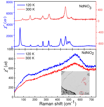
Figure 5 is the Raman spectra of NdNiO3 and NdNiO2 at 300 K and 120 K measured on the sub-millimeter sizable single crystals. The Raman susceptibility deduced from the Raman intensity according to the fluctuation-dissipation theorem, where is the boson factor.
The Raman spectra for NdNiO3 agrees well with the previous measurements on the thin-film single crystal Zaghrioui et al. (2001), implying the high-quality for our bulk NdNiO3 samples. There are more phonon peaks at low temperatures due to the structure distortion during the metal-insulator transition around 200 K in NdNiO3. The dramatically increasing of the phonon intensity is due to the metal-insulator transition in NdNiO3.
The surface contaminant due to the formation of fluorite NdNiOxHy reported in the previous thin-film NdNiO2 Onozuka et al. (2016); Lee et al. (2020) is nearly invisible in our Nd XPS. To remove the possible contaminant, we polish the NdNiO2 single crystal as shown in the inset of Fig. 5. During the synthesis process, some pure nickel metal are embedded in NdNiO2 crystal due to the perotectic phenomenon and forms the dark gray spots (marked by red circles) in the polished surface of NdNiO2. The large light gray area in the SEM image is NdNiO2 as identified by EDX. We perform the Raman scattering measurements on NdNiO2 in the light gray area.
The ideal stoichiometric NdNiO2 has the space group and all the ions can be taken as the inversion center of the lattice. Therefore, there is no Raman active phonon mode in the Raman scattering. There is no sharp phonon modes in the spectra. A broad peak appears at 122 cm-1, independent of the temperature and its origin is unclear. We suspect it comes from the defect due to the off-stoichiometry of NdNiO2.
We attribute the mode at around 550 cm-1 in the Raman susceptibility spectra of NdNiO2 in Fig. 5 to the two-magnon peak in the two-dimensional Heisenberg antiferromagnet. The broadening of the two-magnon response with doping and temperature has been studied theoretically in the cuprate oxides Sugai et al. (1988); Lyons et al. (1988, 1989); Knoll et al. (1990); Sulewski et al. (1990, 1991); Blumberg et al. (1994, 1996); Rübhausen et al. (1997, 1999); Sugai et al. (2003). The two-magnon identifies the strong spin fluctuations in the cuprate even when the long range antiferromagnetic order disappears at high temperatures or upon doping Devereaux and Hackl (2007). In a 2D Heisenberg antiferromagnet, the light in the Raman process flips two nearby spins on the nearest neighboring bond, leaving behind a locally disturbed antiferromagnet with 6 broken exchange bonds in the final state. Therefore the two-magnon has the energy scale at roughly Devereaux and Hackl (2007). We equal to 550 cm-1, and obtain the antiferromagnetic exchange strength meV.
Discussions and Conclusions. – The discovery of the superconductivity in the thin film Nd0.8Sr0.2NiO2 Li et al. (2019) has caught lots of attention, however, the recent report of the bulk Nd1-xSrxNiO2 () reveals an insulating behavior without the presence of the superconductivity Li et al. (2019). The reason of the discrepancy is unclear. We suspect that the Sr substitution of Nd destroys the flat NiO2 plane in the bulk Nd1-xSrxNiO2, probably resulting in the insulating behavior due to the disorder. In the thin film Nd1-xSrxNiO2, due to the strain effect of substrate, the NiO2 plane remains flat after doping.
In our previous theoretical work, we derive the effective Hamiltonian for nickelate oxides Nd1-xSrxNiO2 with the flat NiO2 planes Zhang et al. (2020a). From the the Heyd-Scuseria-Ernzerhof hybrid density functional and the exact diagonalization of the three-band Hubbard model for the Ni5O16 cluster, we find that the undoped NiO2 plane is a Mott-Hubbard insulator and the doped holes primarily locate on Ni sites. The Mott-Hubbard scenario is also stressed in Ref. Jiang et al. (2019a). The physics of the undoped NiO2 plane is described by the two-dimensional Heisenberg antiferromagnet on the square lattice with the exchange strength meV Zhang et al. (2020a).
In summary, we perform core-level X-ray photoemission spectroscopy (XPS) and Raman spectroscopy studies of electronic structures and spin fluctuations in the bulk NdNiO2. We find that the NiO2 planes in NdNiO2 are of Mott-Hubbard type in the ZSA scheme, consistent with Hepting’s electronic structure studies on the thin-film NdNiO2 Hepting et al. (2020). Two-magnon peak in the Raman scattering provides direct evidence for the strong spin-fluctuation in NdNiO2. The peak position determines the antiferromagnetic exchange meV. The present experimental investigation agrees well with our previous theoretical description of the electronic structures of NdNiO2.
In the last, we make a remark on the three-dimensional electron pockets of Nd3+ character in NdNiO2 as demonstrated in Ref. Lee and Pickett (2004); Hepting et al. (2020). The presence of electron pocket changes the hole count in the NiO2 planes, resulting in spin- Ni2+ states in the NiO2 planes even without chemical doping Zhang et al. (2020a). A weakly-interacting three-dimensional 5 metallic state hybridizes with the Ni+ local moments in the NiO2 layers, explaining the resonant inelastic x-ray scattering spectra in LaNiO2 and NdNiO2 Hepting et al. (2020). In our Ni XPS spectra, the main line for NdNiO2 has some weight around 853 eV (Fig. 4). We can’t rule out the minority nickel metal origin for this weight. However, XRD has the similar penetration depth as XPS and detect a tiny nickel peak in our samples. So the 853 eV weight in Fig. 4 for NdNiO2 is likely to be intrinsic and related to the Ni2+ states in the NiO2 planes.
Acknowledgements.
Acknowledgments – J.W.M thanks A. Ng and W. Q. Chen for useful discussions. J.W.M was partially supported by the program for Guangdong Introducing Innovative and Entrepreneurial Teams (No. 2017ZT07C062). This work was also supported by National Science Foundation of China (No. 11774143).References
- Zaanen et al. (1985) J. Zaanen, G. A. Sawatzky, and J. W. Allen, “Band gaps and electronic structure of transition-metal compounds,” Phys. Rev. Lett. 55, 418–421 (1985).
- Medarde (1997) Maria Luisa Medarde, “Structural, magnetic and electronic properties of perovskites (R = rare earth),” Journal of Physics: Condensed Matter 9, 1679–1707 (1997).
- Catalan (2008) G. Catalan, “Progress in perovskite nickelate research,” Phase Transitions 81, 729–749 (2008).
- Middey et al. (2016) S. Middey, J. Chakhalian, P. Mahadevan, J. W. Freeland, A. J. Millis, and D. D. Sarma, “Physics of ultrathin films and heterostructures of rare-earth nickelates,” Annu. Rev. Mater. Res. 46, 305–334 (2016).
- Catalano et al. (2018) S. Catalano, M. Gibert, J. Fowlie, J. Íñiguez, J.-M. Triscone, and J. Kreisel, “Rare-earth nickelates rnio3: thin films and heterostructures,” Reports on Progress in Physics 81, 046501 (2018).
- Crespin et al. (1983) Michel Crespin, Pierre Levitz, and Lucien Gatineau, “Reduced forms of LaNiO3 perovskite. Part 1. – Evidence for new phases: La2Ni2O5 and LaNiO2,” J. Chem. Soc. Faraday Trans. II 79, 1181–1194 (1983).
- Hayward et al. (1999) M. A. Hayward, M. A. Green, M. J. Rosseinsky, and J. Sloan, “Sodium Hydride as a Powerful Reducing Agent for Topotactic Oxide Deintercalation: Synthesis and Characterization of the Nickel(I) Oxide LaNiO2,” J. Am. Chem. Soc. 121, 8843–8854 (1999).
- Hayward and Rosseinsky (2003) M. A. Hayward and M. J. Rosseinsky, “Synthesis of the infinite layer Ni(I) phase NdNiO2+x by low temperature reduction of NdNiO3 with sodium hydride,” Solid State Sciences 5, 839–850 (2003).
- Anisimov et al. (1999) V. I. Anisimov, D. Bukhvalov, and T. M. Rice, “Electronic structure of possible nickelate analogs to the cuprates,” Phys. Rev. B 59, 7901–7906 (1999).
- Lee and Pickett (2004) K.-W. Lee and W. E. Pickett, “Infinite-layer LaNiO2: Ni1+ is not Cu2+,” Phys. Rev. B 70, 165109 (2004).
- Kawai et al. (2009) Masanori Kawai, Satoru Inoue, Masaichiro Mizumaki, Naomi Kawamura, Noriya Ichikawa, and Yuichi Shimakawa, “Reversible changes of epitaxial thin films from perovskite LaNiO3 to infinite-layer structure LaNiO2,” Appl. Phys. Lett. 94, 082102 (2009).
- Kaneko et al. (2009) D. Kaneko, K. Yamagishi, A. Tsukada, T. Manabe, and M. Naito, “Synthesis of infinite-layer LaNiO2 films by metal organic decomposition,” Physica C: Superconductivity 469, 936–939 (2009).
- Kawai et al. (2010) Masanori Kawai, Kazuya Matsumoto, Noriya Ichikawa, Masaichiro Mizumaki, Osami Sakata, Naomi Kawamura, Shigeru Kimura, and Yuichi Shimakawa, “Orientation Change of an Infinite-Layer Structure LaNiO2 Epitaxial Thin Film by Annealing with CaH2,” Crystal Growth & Design 10, 2044–2046 (2010).
- Ikeda et al. (2013) Ai Ikeda, Takaaki Manabe, and Michio Naito, “Improved conductivity of infinite-layer LaNiO2 thin films by metal organic decomposition,” Physica C: Superconductivity 495, 134–140 (2013).
- Ikeda et al. (2016) Ai Ikeda, Yoshiharu Krockenberger, Hiroshi Irie, Michio Naito, and Hideki Yamamoto, “Direct observation of infinite NiO2 planes in LaNiO2 films,” Applied Physics Express 9, 061101 (2016).
- Li et al. (2019) Danfeng Li, Kyuho Lee, Bai Yang Wang, Motoki Osada, Samuel Crossley, Hye Ryoung Lee, Yi Cui, Yasuyuki Hikita, and Harold Y. Hwang, “Superconductivity in an infinite-layer nickelate,” Nature 572, 624–627 (2019).
- Hepting et al. (2020) M. Hepting, D. Li, C. J. Jia, H. Lu, E. Paris, Y. Tseng, X. Feng, M. Osada, E. Been, Y. Hikita, Y.-D. Chuang, Z. Hussain, K. J. Zhou, A. Nag, M. Garcia-Fernandez, M. Rossi, H. Y. Huang, D. J. Huang, Z. X. Shen, T. Schmitt, H. Y. Hwang, B. Moritz, J. Zaanen, T. P. Devereaux, and W. S. Lee, “Electronic structure of the parent compound of superconducting infinite-layer nickelates,” Nature Materials (2020).
- Bednorz and Müller (1986) J. G. Bednorz and K. A. Müller, “Possible highTc superconductivity in the Ba-La-Cu-O system,” Z. Physik B Condensed Matter 64, 189–193 (1986).
- Botana and Norman (2020) AS Botana and MR Norman, “Similarities and Differences between LaNiO2 and CaCuO2 and Implications for Superconductivity,” Phys. Rev. X 10, 011024 (2020).
- Sakakibara et al. (2019) Hirofumi Sakakibara, Hidetomo Usui, Katsuhiro Suzuki, Takao Kotani, Hideo Aoki, and Kazuhiko Kuroki, “Model construction and a possibility of cuprate-like pairing in a new d9 nickelate superconductor (Nd,Sr)NiO2,” (2019), arXiv:1909.00060 [cond-mat.supr-con] .
- Jiang et al. (2019a) Mi Jiang, Mona Berciu, and George A. Sawatzky, “Doped holes in NdNiO2 and high- cuprates show little similarity,” (2019a), arXiv:1909.02557 [cond-mat.supr-con] .
- Wu et al. (2020) Xianxin Wu, Domenico Di Sante, Tilman Schwemmer, Werner Hanke, Harold Y Hwang, Srinivas Raghu, and Ronny Thomale, “Robust -wave superconductivity of infinite-layer nickelates,” Phy. Rev. B 101, 060504 (2020).
- Nomura et al. (2019) Yusuke Nomura, Motoaki Hirayama, Terumasa Tadano, Yoshihide Yoshimoto, Kazuma Nakamura, and Ryotaro Arita, “Formation of a two-dimensional single-component correlated electron system and band engineering in the nickelate superconductor ndnio 2,” Physical Review B 100, 205138 (2019).
- Gao et al. (2019) Jiacheng Gao, Zhijun Wang, Chen Fang, and Hongming Weng, “Electronic structures and topological properties in nickelates NinO2n+2,” (2019), arXiv:1909.04657 [cond-mat.mtrl-sci] .
- Zhang et al. (2020a) Hu Zhang, Lipeng Jin, Shanmin Wang, Bin Xi, Xingqiang Shi, Fei Ye, and Jia-Wei Mei, “Effective Hamiltonian for nickelate oxides Nd1-xSrxNiO2,” Phys. Rev. Research 2, 013214 (2020a).
- Ryee et al. (2020) Siheon Ryee, Hongkee Yoon, Taek Jung Kim, Min Yong Jeong, and Myung Joon Han, “Induced magnetic two-dimensionality by hole doping in the superconducting infinite-layer nickelate Nd1-xSrxNiO2,” Phys. Rev. B 101, 064513 (2020).
- Zhang et al. (2020b) Guang-Ming Zhang, Yi-feng Yang, and Fu-Chun Zhang, “Self-doped mott insulator for parent compounds of nickelate superconductors,” Phys. Rev. B 101, 020501 (2020b).
- Zhang and Vishwanath (2019) Ya-Hui Zhang and Ashvin Vishwanath, “Type II model in superconducting nickelate Nd1-xSrxNiO2,” (2019), arXiv:1909.12865 [cond-mat.str-el] .
- Jiang et al. (2019b) Peiheng Jiang, Liang Si, Zhaoliang Liao, and Zhicheng Zhong, “Electronic structure of rare-earth infinite-layer ,” Phys. Rev. B 100, 201106 (2019b).
- Hu and Wu (2019) Lun-Hui Hu and Congjun Wu, “Two-band model for magnetism and superconductivity in nickelates,” Phys. Rev. Research 1, 032046 (2019).
- Chang et al. (2019) Jun Chang, Jize Zhao, and Yang Ding, “Hund-heisenberg model in superconducting infinite-layer nickelates,” (2019), arXiv:1911.12731 [cond-mat.supr-con] .
- Werner and Hoshino (2020) Philipp Werner and Shintaro Hoshino, “Nickelate superconductors: Multiorbital nature and spin freezing,” Phys. Rev. B 101, 041104 (2020).
- Liu et al. (2019) Zhao Liu, Zhi Ren, W. Zhu, Z. F. Wang, and Jinlong Yang, “Electronic and magnetic structure of infinite-layer : Trace of antiferromagnetic metal,” (2019), arXiv:1912.01332 [cond-mat.str-el] .
- Choi et al. (2020) Mi-Young Choi, Kwan-Woo Lee, and Warren E. Pickett, “Role of 4 states in infinite-layer NdNiO2,” Physical Review B 101 (2020), 10.1103/physrevb.101.020503.
- Karp et al. (2020) Jonathan Karp, Antia S. Botana, Michael R. Norman, Hyowon Park, Manuel Zingl, and Andrew Millis, “Many-body Electronic Structure of NdNiO2 and CaCuO2,” arXiv e-prints , arXiv:2001.06441 (2020), arXiv:2001.06441 [cond-mat.str-el] .
- Li et al. (2020) Danfeng Li, Bai Yang Wang, Kyuho Lee, Shannon P. Harvey, Motoki Osada, Berit H. Goodge, Lena F. Kourkoutis, and Harold Y. Hwang, “Superconducting Dome in Nd1-xSrxNiO2 Infinite Layer Films,” arXiv e-prints , arXiv:2003.08506 (2020), arXiv:2003.08506 [cond-mat.supr-con] .
- Zhang and Rice (1988) F. C. Zhang and T. M. Rice, “Effective Hamiltonian for the superconducting Cu oxides,” Phys. Rev. B 37, 3759–3761 (1988).
- Fujimori et al. (1987) A. Fujimori, E. Takayama-Muromachi, Y. Uchida, and B. Okai, “Spectroscopic evidence for strongly correlated electronic states in La-Sr-Cu and Y-Ba-Cu oxides,” Phys. Rev. B 35, 8814–8817 (1987).
- Shen et al. (1987) Zhi-xun Shen, J. W. Allen, J. J. Yeh, J. S. Kang, W. Ellis, W. Spicer, I. Lindau, M. B. Maple, Y. D. Dalichaouch, M. S. Torikachvili, J. Z. Sun, and T. H. Geballe, “Anderson hamiltonian description of the experimental electronic structure and magnetic interactions of copper oxide superconductors,” Phys. Rev. B 36, 8414–8428 (1987).
- van Veenendaal and Sawatzky (1993) M. A. van Veenendaal and G. A. Sawatzky, “Nonlocal screening effects in 2p x-ray photoemission spectroscopy core-level line shapes of transition metal compounds,” Phys. Rev. Lett. 70, 2459–2462 (1993).
- van Veenendaal (2006) Michel van Veenendaal, “Competition between screening channels in core-level x-ray photoemission as a probe of changes in the ground-state properties of transition-metal compounds,” Phys. Rev. B 74, 085118 (2006).
- Ghiasi et al. (2019) Mahnaz Ghiasi, Atsushi Hariki, Mathias Winder, Jan Kuneš, Anna Regoutz, Tien-Lin Lee, Yongfeng Hu, Jean-Pascal Rueff, and Frank M. F. de Groot, “Charge-transfer effect in hard x-ray and photoemission spectra: and cluster-model analysis,” Phys. Rev. B 100, 075146 (2019).
- Anderson (1987) Philip W Anderson, “The Resonating Valence Bond State in La2CuO4 and Superconductivity.” Science 235, 1196–8 (1987).
- Lee et al. (2006) Patrick A Lee, Naoto Nagaosa, and Xiao-Gang Wen, “Doping a Mott insulator: Physics of high-temperature superconductivity,” Rev. Mod. Phys. 78, 17 (2006).
- Devereaux and Hackl (2007) Thomas P. Devereaux and Rudi Hackl, “Inelastic light scattering from correlated electrons,” Rev. Mod. Phys. 79, 175–233 (2007).
- Sugai et al. (1988) Shunji Sugai, Shin-ichi Shamoto, and Masatoshi Sato, “Two-magnon Raman scattering in (,” Phys. Rev. B 38, 6436–6439 (1988).
- Lyons et al. (1988) K. B. Lyons, P. A. Fleury, J. P. Remeika, A. S. Cooper, and T. J. Negran, “Dynamics of spin fluctuations in lanthanum cuprate,” Phys. Rev. B 37, 2353–2356 (1988).
- Lyons et al. (1989) K. B. Lyons, P. E. Sulewski, P. A. Fleury, H. L. Carter, A. S. Cooper, G. P. Espinosa, Z. Fisk, and S.-W. Cheong, “High-energy spin and charge excitations in ,” Phys. Rev. B 39, 9693–9696 (1989).
- Knoll et al. (1990) P. Knoll, C. Thomsen, M. Cardona, and P. Murugaraj, “Temperature-dependent lifetime of spin excitations in R (R=Eu, Y),” Phys. Rev. B 42, 4842–4845 (1990).
- Sulewski et al. (1990) P. E. Sulewski, P. A. Fleury, K. B. Lyons, S-W. Cheong, and Z. Fisk, “Light scattering from quantum spin fluctuations in (R=La, Nd, Sm),” Phys. Rev. B 41, 225–230 (1990).
- Sulewski et al. (1991) P. E. Sulewski, P. A. Fleury, K. B. Lyons, and S-W. Cheong, “Observation of chiral spin fluctuations in insulating planar cuprates,” Phys. Rev. Lett. 67, 3864–3867 (1991).
- Blumberg et al. (1994) G. Blumberg, R. Liu, M. V. Klein, W. C. Lee, D. M. Ginsberg, C. Gu, B. W. Veal, and B. Dabrowski, “Two-magnon raman scattering in cuprate superconductors: Evolution of magnetic fluctuations with doping,” Phys. Rev. B 49, 13295–13298 (1994).
- Blumberg et al. (1996) G. Blumberg, P. Abbamonte, M. V. Klein, W. C. Lee, D. M. Ginsberg, L. L. Miller, and A. Zibold, “Resonant two-magnon raman scattering in cuprate antiferromagnetic insulators,” Phys. Rev. B 53, R11930–R11933 (1996).
- Rübhausen et al. (1997) M. Rübhausen, C. T. Rieck, N. Dieckmann, K.-O. Subke, A. Bock, and U. Merkt, “Inelastic light scattering at high-energy excitations in doped cuprate superconductors,” Phys. Rev. B 56, 14797–14808 (1997).
- Rübhausen et al. (1999) M. Rübhausen, O. A. Hammerstein, A. Bock, U. Merkt, C. T. Rieck, P. Guptasarma, D. G. Hinks, and M. V. Klein, “Doping Dependence of the Electronic Interactions in Bi-2212 Cuprate Superconductors: Doped Antiferromagnets or Antiferromagnetic Fermi Liquids?” Phys. Rev. Lett. 82, 5349–5352 (1999).
- Sugai et al. (2003) S. Sugai, H. Suzuki, Y. Takayanagi, T. Hosokawa, and N. Hayamizu, “Carrier-density-dependent momentum shift of the coherent peak and the lo phonon mode in p-type high- superconductors,” Phys. Rev. B 68, 184504 (2003).
- Vassiliou et al. (1989) John K Vassiliou, Marc Hornbostel, Robin Ziebarth, and Francis J Disalvo, “Synthesis and properties of NdNiO3 prepared by low-temperature methods,” Journal of Solid State Chemistry 81, 208–216 (1989).
- Li et al. (2019) Qing Li, Chengping He, Jin Si, Xiyu Zhu, Yue Zhang, and Hai-Hu Wen, “Absence of superconductivity in bulk Nd1-xSrxNiO2 (x = 0.2, 0.4),” arXiv e-prints , arXiv:1911.02420 (2019), arXiv:1911.02420 [cond-mat.supr-con] .
- Rodríguez-Carvajal (1993) Juan Rodríguez-Carvajal, “Recent advances in magnetic structure determination by neutron powder diffraction,” Physica B: Condensed Matter 192, 55 – 69 (1993).
- García-Muñoz et al. (2009) J. L. García-Muñoz, M. A. G. Aranda, J. A. Alonso, and M. J. Martínez-Lope, “Structure and charge order in the antiferromagnetic band-insulating phase of ,” Phys. Rev. B 79, 134432 (2009).
- Toby (2001) Brian H. Toby, “EXPGUI, a graphical user interface for GSAS,” Journal of Applied Crystallography 34, 210–213 (2001).
- De Groot and Kotani (2008) Frank De Groot and Akio Kotani, Core level spectroscopy of solids (CRC press, 2008).
- Van Veenendaal (2015) Michel Van Veenendaal, Theory of Inelastic Scattering and Absorption of X-rays (Cambridge University Press, 2015).
- van der Laan et al. (1981) G. van der Laan, C. Westra, C. Haas, and G. A. Sawatzky, “Satellite structure in photoelectron and auger spectra of copper dihalides,” Phys. Rev. B 23, 4369–4380 (1981).
- Gunnarsson and Schönhammer (1983) O. Gunnarsson and K. Schönhammer, “Photoemission from ce compounds: Exact model calculation in the limit of large degeneracy,” Phys. Rev. Lett. 50, 604–607 (1983).
- Zaanen et al. (1986) J. Zaanen, C. Westra, and G. A. Sawatzky, “Determination of the electronic structure of transition-metal compounds: 2p x-ray photoemission spectroscopy of the nickel dihalides,” Phys. Rev. B 33, 8060–8073 (1986).
- Bocquet et al. (1992) A. E. Bocquet, T. Mizokawa, T. Saitoh, H. Namatame, and A. Fujimori, “Electronic structure of -transition-metal compounds by analysis of the core-level photoemission spectra,” Phys. Rev. B 46, 3771–3784 (1992).
- Sarma and Chainani (1990) D. D. Sarma and A. Chainani, “Systematics in the oxygen 1s core-level photoemission spectra from metal oxides: Model calculations,” Phys. Rev. B 41, 6688–6691 (1990).
- Biesinger et al. (2009) Mark C Biesinger, Brad P Payne, Leo WM Lau, Andrea Gerson, and Roger St C Smart, “X-ray photoelectron spectroscopic chemical state quantification of mixed nickel metal, oxide and hydroxide systems,” Surface and Interface Analysis: An International Journal devoted to the development and application of techniques for the analysis of surfaces, interfaces and thin films 41, 324–332 (2009).
- Hagelin-Weaver et al. (2004) Helena AE Hagelin-Weaver, Jason F Weaver, Gar B Hoflund, and Ghaleb N Salaita, “Electron energy loss spectroscopic investigation of Ni metal and NiO before and after surface reduction by Ar+ bombardment,” Journal of electron spectroscopy and related phenomena 134, 139–171 (2004).
- Payne et al. (2009) BP Payne, MC Biesinger, and NS McIntyre, “The study of polycrystalline nickel metal oxidation by water vapour,” Journal of Electron Spectroscopy and Related Phenomena 175, 55–65 (2009).
- Benndorf et al. (1982) C Benndorf, Ch Nöbl, and F Thieme, “Interaction of H2O with a clean and oxygen precovered Ni (110) surface studied by XPS,” Surface Science 121, 249–259 (1982).
- Carley et al. (1983) AF Carley, S Rassias, and MW Roberts, “The specificity of surface oxygen in the activation of adsorbed water at metal surfaces,” Surface science 135, 35–51 (1983).
- Weaver et al. (1988) J. H. Weaver, H. M. Meyer, T. J. Wagener, D. M. Hill, Y. Gao, D. Peterson, Z. Fisk, and A. J. Arko, “Valence bands, oxygen in planes and chains, and surface changes for single crystals of and M (M=Pr,Nd,Eu,Gd),” Phys. Rev. B 38, 4668–4676 (1988).
- Johnston et al. (2014) Steve Johnston, Anamitra Mukherjee, Ilya Elfimov, Mona Berciu, and George A. Sawatzky, “Charge disproportionation without charge transfer in the rare-earth-element nickelates as a possible mechanism for the metal-insulator transition,” Phys. Rev. Lett. 112, 106404 (2014).
- Mizokawa et al. (1991) T Mizokawa, H Namatame, A Fujimori, K Akeyama, Hiroshi Kondoh, H Kuroda, and N Kosugi, “Origin of the band gap in the negative charge-transfer-energy compound NaCuO2,” Phys. Rev. Lett. 67, 1638 (1991).
- Mizokawa et al. (1995) T Mizokawa, A Fujimori, T Arima, Y Tokura, N Mōri, and Jun Akimitsu, “Electronic structure of prnio 3 studied by photoemission and x-ray-absorption spectroscopy: Band gap and orbital ordering,” Physical Review B 52, 13865 (1995).
- Abbate et al. (2002) M Abbate, G Zampieri, F Prado, A Caneiro, JM Gonzalez-Calbet, and M Vallet-Regi, “Electronic structure and metal-insulator transition in LaNiO3-δ,” Physical Review B 65, 155101 (2002).
- Bisogni et al. (2016) Valentina Bisogni, Sara Catalano, Robert J. Green, Marta Gibert, Raoul Scherwitzl, Yaobo Huang, Vladimir N. Strocov, Pavlo Zubko, Shadi Balandeh, Jean-Marc Triscone, George Sawatzky, and Thorsten Schmitt, “Ground-state oxygen holes and the metal-insulator transition in the negative charge-transfer rare-earth nickelates,” Nature Communications 7, 13017 (2016).
- Zaghrioui et al. (2001) M Zaghrioui, A Bulou, P Lacorre, and P Laffez, “Electron diffraction and Raman scattering evidence of a symmetry breaking at the metal-insulator transition of NdNiO3,” Phys. Rev. B 64, 081102 (2001).
- Onozuka et al. (2016) T. Onozuka, A. Chikamatsu, T. Katayama, T. Fukumura, and T. Hasegawa, “Formation of defect-fluorite structured ndnioxhy epitaxial thin films via a soft chemical route from ndnio3 precursors,” Dalton Trans. 45, 12114–12118 (2016).
- Lee et al. (2020) Kyuho Lee, Berit H. Goodge, Danfeng Li, Motoki Osada, Bai Yang Wang, Yi Cui, Lena F. Kourkoutis, and Harold Y. Hwang, “Aspects of the Synthesis of Thin Film Superconducting Infinite-Layer Nickelates,” arXiv e-prints , arXiv:2002.07749 (2020), arXiv:2002.07749 [cond-mat.mtrl-sci] .