Sensing individual nuclear spins with a single rare-earth electron spin
Sensing individual nuclear spins with a single rare-earth electron spin
Abstract
Rare-earth related electron spins in crystalline hosts are unique material systems, as they can potentially provide a direct interface between telecom band photons and long-lived spin quantum bits. Specifically, their optically accessible electron spins in solids interacting with nuclear spins in their environment are valuable quantum memory resources. Detection of nearby individual nuclear spins, so far exclusively shown for few dilute nuclear spin bath host systems such as the NV center in diamond or the silicon vacancy in silicon carbide, remained an open challenge for rare-earths in their host materials, which typically exhibit dense nuclear spin baths. Here, we present the electron spin spectroscopy of single Ce3+ ions in a yttrium orthosilicate host, featuring a coherence time of s. This coherent interaction time is sufficiently long to isolate proximal 89Y nuclear spins from the nuclear spin bath of 89Y. Furthermore, it allows for the detection of a single nearby 29Si nuclear spin, native to the host material with ~5 % abundance. This study opens the door to quantum memory applications in rare-earth ion related systems based on coupled environmental nuclear spins, potentially useful for quantum error correction schemes.
Hybrid quantum systems, consisting of a read-out electron spin and long-lived nuclear spins, have demonstrated remarkable properties for quantum memory applications Morton et al. (2008); Maurer et al. (2012); Awschalom et al. (2018). At the same time single spins enable active quantum processing used in e.g. quantum error correction Waldherr et al. (2014). Implementing them in scalable quantum networks based on single rare-earth ions (REI) doped in solids potentially combines long distance entanglement distribution via single telecom band photonsDibos et al. (2018) with error corrected long-lived quantum memories.
Based on the efficient isolation of REI’s electrons, their narrow and stable optical and spin levels have been used for demonstration of storage and retrieval of single photons De Riedmatten and Afzelius (2015) and exceptional coherence times Zhong et al. (2015), rendering them particularly suitable for quantum repeater protocols. With additional control over single rare-earth ions, however, these capabilities can be extended to high-fidelity spin readout and generation of entanglement of rare-earth electrons and nuclei in a scalable fashionAwschalom et al. (2018). Consequently, an increasing number of REI are isolated as single emitters Kolesov et al. (2012); Yin et al. (2013); Kolesov et al. (2013); Zhong et al. (2018); Dibos et al. (2018). Based on ancillary electron spins of these single emitters, sensing of nuclear spins Zhao et al. (2012a); Kolkowitz et al. (2012); Taminiau et al. (2012); Car et al. (2018); Zhong et al. (2019) is an important next step for REI based quantum network applications. So far, only dilute nuclear spin bath host materials such as diamond Childress et al. (2006), silicon carbide Nagy et al. (2019) and silicon Pla et al. (2013) were successfully used for detection of individual nuclear spins.
In this study, we use the yttrium orthosilicate (Y2SiO5,YSO) crystal to investigate the nulcear environment of individual Ce3+ electron spins, which simultaneously act as a proxy for other REI species, owing to their interchangable doping into yttrium containing solids. We demonstrate spin initialization and coherent manipulation of Ce3+ electron spins in a YSO host crystal. Surprisingly we were able to sense individual dipolar coupled 29Si nuclear spins despite the strong yttrium spin bath. 29Si signal is distilled furthermore with basic decoupling sequences. Signatures of yttrium nuclear spins also reveal dipolar coupling with the nearby Ce3+ superimposed on the yttrium spin bath.
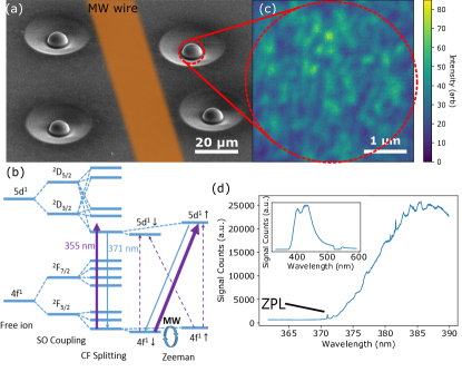
Trivalent cerium substitutes Y3+ in 95 % of the cases at the 7-oxygen-coordinated site of the YSO crystal with a symmetry Pidol et al. (2006). The remaining 5 % of Ce3+ ions substituting Y3+ in the 6-oxygen-coordinated site of the crystal can be neclegted from further considerations due to different optical (red shifted) and magnetic (different g-tensor) properties Pidol et al. (2006); Drozdowski et al. (2004). Individual Ce3+ ions were identified in an ultra-pure YSO crystal using laser scanning confocal microscopy. The experimental setup of the microscope is described in detail in the supplementary material sup . To improve the collection efficiency and spatial resolution of the microscope, solid immersion lenses (SIL) were fabricated on the surface of the sample by focused ion beam milling. A scanning electron microscopy (SEM) image of the sample with milled SILs and an indicated wire used for microwave (MW) spin manipulation is shown in Fig. 1a. A picosecond pulsed laser at 355 nm wavelength was used to off-resonantly excite Ce3+ ions from the ground state into the excited band based on their phonon-assisted absorption band Drozdowski et al. (2004). The corresponding energy level structure of Ce3+ in YSO is shown in Fig. 1b. For the purpose of acquiring a fluorescence spectrum, emission was collected from a 0.01% doped Ce3+:YSO crystal at K (inset Fig. 1d). A high resolution spectrum of Ce3+:YSO, shown in Fig. 1d, reveals the zero-phonon line at 371 nm, characteristic for Ce3+ ion fluorescence at cryogenic temperatures Yan et al. (2013). Performing confocal microscopy with the pulsed 355 nm laser on the SILs, however, allows for resolving individual Ce3+ ions. A typical laser scanning fluorescence image of optically resolved Ce3+ ions located in the focus of a SIL is shown in Fig. 1c.
Even in ultra-pure crystals (from Scientific Materials), cerium is an unavoidable native impurity for yttrium-based hosts and the estimated residual density of 0.3 ppb in our crystal is the main contribution to background signal and can result in more than one Ce3+ ion to be probed within the focal volume. In principle, stimulated emission depletion (STED) based superresolution microscopy is available for Ce3+:YSO (see supplementary material sup ) Kolesov et al. (2018), however the spin initialization and readout was found to be prevented by the high power depletion laser used in the experiment.
In a magnetic field parallel to the optical excitation beam, optical transitions between and spin doublets show different relative strengths under circularly polarized excitation Kolesov et al. (2013). While the spin-flip transition exhibits the strongest optical dipole moment out of the four possible transitions under circularly polarized excitation (indicated in Fig. 1b), the radiative decay originating from the excited level ends up in both ground state spin levels with equal probability. A repeated excitation of the selectively driven spin-flip transition eventually results in a polarized ground state spin level. When applying the optical polarization procedure, the spin is pumped into the optically ’dark’ stateSiyushev et al. (2014), which results in a reduced fluorescence signal. MW radiation resonant with the ground state spin transition can then be used to flip the optically polarized spin. This results in an increased fluorescence signal, which allows for optically detected magnetic resonance (ODMR) measurements on individual cerium ions.
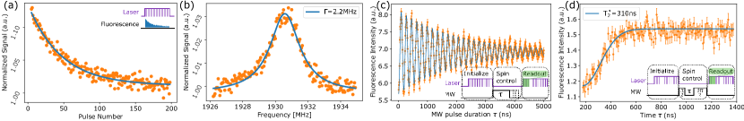
Initialization measurements, shown in Fig. 2a., capture the dynamic evolution of the fluorescence signal as a function of the laser pulse number. Starting with a thermal polarization, the fluorescence signal drops with increasing number of laser pulses because of optical pumping of the spin. Initialization fidelities range between 5% and 15%, depending on the individual Ce3+ ion under investigation. The relatively high background of densely packed Ce3+ ions in the sample can contribute to measuring a decreased fidelity.
In our experiments, a magnetic field with a strength of 970 Gauss was applied parallel to the optical beam and the -axis of the YSO crystal, for which Ce3+ has a -factor of and a magnetic resonance frequency of 1930.5 MHzPidol et al. (2006). MW structures created in proximity of SILs were then used to sweep the MW radiation frequency in order to observe ODMR spectra of individual Ce3+ ions. A typical spectrum is shown in Fig. 2b and exhibits a linewidth of 2-3 MHz, slightly different for different cerium ions.
Coherent Ce3+ spin manipulation is demonstrated in a measurement of spin Rabi oscillations under strong MW driving, shown in Fig. 2c. A typical sequence for such a Rabi measurement contains spin initialization, control and readout, as shown in the inset of Fig. 2c. An exponantially decaying sine function was fitted to the Rabi measurement signal, revealing a Rabi frequency of 5.6 MHz and a decay time of 2 s.
A free induction decay (FID) experiment is capable to reveal the electron spin coherence in the thermal noise of the characteristic nuclear spin bath of YSO, featuring 89Y and 29Si nuclear spins. Fig. 2d presents a typical FID of Ce3+ and quantifies the inhomogenous broadening of Ce3+ spin transition to ns, by fitting with a Gaussian. These dephasing times are in good agreement with ODMR linewidth measurements. The main contribution to inhomogenous broadening may emerge from the nuclear spin bath and can also come from short-pulsed optical excitation of Ce3+, causing ionization of electron traps nearby the electron spin, which induce charge fluctuations capable of stark-shifting the resonance.
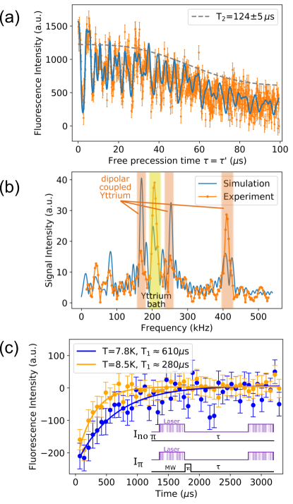
The Hahn spin echo sequence decouples the spin from slow (compared to ) changes in the environment and allows for more detailed spin spectroscopy of the nuclear spin environment of Ce3+ electron spins. A respresentative Hahn echo measurement on Ce3+ is shown in Fig. 3a. We observe periodic revivals related to yttrium ions in the crystal, which have 100% abundance of nuclear spin with magnetic moment , with as the nuclear magneton. The overall decaying signal corresponds to a decoherene time of s and was fitted to Childress et al. (2006). Based on the cluster-correlation expansion method Zhao et al. (2012b), the coherence of the electron spin in an interacting bath is simulated and plotted as blue line in Fig. 3a. For the Ce3+ electron spin located in a complex nuclear environment such as the YSO matrix, the simulated behaviour of its coherence under spin echo control agrees strikingly well with our experimental results. Details of the simulations are described in the supplementary material sup . The Fast Fourier Transform (FFT) of the Hahn echo signal (see Fig. 3b) reveals two particular facts. Firstly, yttrium bath related signatures, found at the Lamor precession frequency kHz, similarly known from single NV centers in diamond Childress et al. (2006). Additionally, we find frequency components related to weakly coupled yttrium nuclear spins based on magnetic dipole interaction with the electron spin. The measured hyperfine coupling strengths are in good agreement with simulated splittings (in the range between 50 kHz and 120 kHz).
We measured the lifetime of the spin state after optical initialization by reading out the spin state after a variable waiting time . The spin relaxation measurements are shown in Fig. 3c and reveal s at a temperature of K, measured at the heat exchanger of our cold-finger cryostat. At an indicated temperature of K, the spin lattice relaxation measurement yields s. By comparing our measured values with EPR based values Kurkin and Chernov (1980), we can identify the actual sample temperatures to be approximatly 4 K higher than measured on the heat exchanger, because of insufficient cooling power.
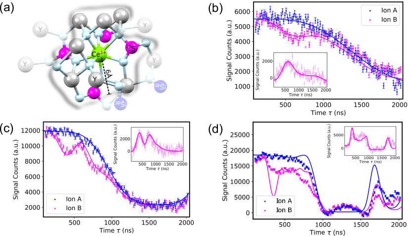
Using confocal microscopy, we can study spatially resolved Ce3+ ions and their unique environment. For each silicon ion in the crystal lattice, there is a 4.7 % natural abundance of 29Si isotope with a nuclear spin and . For a Ce3+ ion under investigation, this leads to a chance of approximately 20 % to have a 29Si as nearest neighbor. At the nearest neighbor location, the close distance (Å) between electron spin and 29Si nuclear spin leads to a detectable hyperfine coupling based on magnetic dipole interaction, superimposed on the hyperfine interaction with closeby yttrium spins and the yttrium bath (schematically shown in Fig. 4a).
Dynamic decoupling (DD) of the Ce3+ spin from the nuclear bath allows to extract 29Si related
signatures. Carr-Purcell-Meiboom-Gill (CPMG) control sequences were used to acquire noise spectra
for two different Ce3+ ions, as shown in Fig. 4(b)-(d).
The center of the
coherence dip (in Fig. 4b and inset) at ns,
corresponds to a revival time of
. matches well with the
gyromagnetic ratio
MHz/T, thus confirming the nearby 29Si nuclear spin.
Specifically, CPMG- decoupling sequences were used, with denoting the number of
pulses used in the sequences.
The bandwidth depends on the number of pulses and scales approximately with at frequency .
Zhao et al. (2011).
With an increasing number of pulses, the noise filter function
causes the 29Si signature to be split in coherence dips. This
can be seen in the insets of Fig. 4(b)-(d), where the 29Si signal is isolated by plotting
the difference between signals from the two Ce3+ ions under investigation.
Solid lines in Fig. 4 are simulated noise spectra of Ce3+ ions located in
a specific nuclear spin environment.
In order to account for differences in the depth of coherence dips between experiment
and simulation, we phenomenologically add a relaxation and dephasing
mechanism to the dynamics of (see sup ).
One possible reason
for a reduced depth of the coherence dip in the experiment is
external noise for proximal . Broadening mechanisms can be related to
magnetic field fluctuations introduced by MW manipulation of Ce3+,
the optical initialization as described for the NV center system Wang and Yang (2015)
or residual RE Kramers ion impurities in the crystal
(such as Er3+, Gd3+,), acting as a noise source on a short timescale.
Based on our simulations, we can localize a coupled
29Si nuclear spin within the nearest neighbor position in the lattice (”ion B”, magenta).
Furthermore,
the comparison spectrum without 29Si nuclear spin signatures (”ion A”, blue) reveals information
about the absence of 29Si within 6 Å distance from the Ce3+ ion. The probability
to find a Ce3+ ion with the same nuclear spin environment as ”ion A” in YSO is
%.
In conclusion, we show coherent control of an individual Ce3+ electron spin in a YSO matrix. Using spin decoupling techniques, our spin spectroscopy reveals the single REI electron spin to be dipolar coupled to nearby nuclear spins. Based on high density of yttrium in the host crystal, the future challenge is to distinguish spectrally between individual coupled nulcear spins. Carefully designed DD sequences can improve detection of nuclear spin signals, which tend to be submerged by the noisy spin bath Ma et al. (2015). 29Si nuclear spin sensing was demonstrated, for 29Si being located within the nearest neighbor shell.
Nuclear spins (either nuclear spin or yttrium nuclear spin) in proximity to the Ce3+ electron spin are quantum resources for quantum memory protocols. By establishing polarization transfer techniques, for example based on Hartmann-Hahn double resonance techniquesLondon et al. (2013), single 29Si nuclear spin could be initialized and potentially used as memory. A key concern is the coherence time of nuclear spins in this context. Decoherence caused by the nuclear spin bath will set the ultimate limitation for the coherence time. Due to weak dipolar interaction between nuclear spins in YSO compared to the Zeeman energy at applied magnetic fields, only the pure-dephasing interaction can have significant effect on the decoherence Zhao et al. (2012b). The characteristic decoherence time scale can be estimated by the nuclear spin dipolar interaction and gives for nuclear spin and for yttrium nuclear spin.
Finally, this work motivates the realization of controllable multispin quantum registers based on single REIs embedded in the YSO matrix. Access to local nodes based on environmental spins, as demonstrated in Waldherr et al. (2014), provides functionality of quantum memories, such as error correction. Furthermore, presented findings are applicable to other Kramers ions doped into YSO, such as erbium, for which coherent spin control and readout was demonstrated recently Raha et al. (2019).
R.K. acknowledges financial support by the DFG (Grant No. KO4999/3-1) and R.K. and J.W. acknowledge financial support by the FET-Flagship Project SQUARE, the EU via SMeL and QIA as well as the DFG via FOR 2724. N.Z. acknowledges financial support by NSFC (Grant No. 11534002) and NSAF (Grant No. U1530401 and Grant No. U1730449).
Supplementary Information
Laser Scanning Confocal Microscope Setup
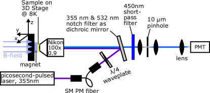
The experiment was carried out in a cold-finger cryostat (CryoVac, KONTI-CRYOSTAT TYPE MICRO), where the sample is mounted on top of a permament magnet, which is mounted on the cold-finger (sketch shown in Fig. 5). The cold-finger is connected to the heat-exchanger, where also the temperature measurement is picked up. Due to low heat conductivity of the magnet between sample and cold-finger, the cooling power is insufficient for thermalizing the YSO sample to the indicated temperature of K, but rather to K. For rough positioning, the cold-finger is mounted on a 3D stage covering 10mm x 10mm x 10mm. To obtain high resolution fluorescence images, the objective lens, mounted on a 3D piezo stage (NPoint, NPXY100Z25-264) in the cryostat vacuum chamber, can be positioned throughout 100m x 100m x 25m with nanometer resolution.
In order to obtain a picosecond-pulsed laser at around 355 nm wavelength, we used a dye laser cavity, which was synchronously pumped by a 532 nm picosecond pulsed Vanguard laser (Spectra-Physics) with 2W output power and 86 MHz repetition rate. The pumped Pyridin 1 dye radiation emitting at 710 nm was extracted from the laser cavity by an intra-cavity, externally triggerable Quartz AOM. With a BBO doubling crystal, we converted the pulsed 710 nm radiation into 355 nm and sent it through a single-mode polarization maintaining (Thorlabs, PANDA PM-S350-HP) fiber for geometric mode cleaning. The linearly polarized light after the output of the fiber was then sent through a waveplate. It was found, that optical polarization fidelity of Ce3+ was not maximum at 45∘ tilt-angle of the waveplate, corresponding to perfectly circularly polarized light, but rather at slightly smaller tilt-angles of 40∘, corresponding to slightly elliptically polarized light. This can originate from birefringence of the YSO crystal and also the 355 nm notch filter, used as a dichroic mirror in the confocal microscope setup, both capable to distort the polarization of light, which would have to be compensated. The fluorescence of Ce3+ was filtered with a 450 nm shortpass filter, in order to reject parasitic emission from impurities found in the YSO crystal, potentially originating from other RE ion species. Additionally, flourescence was filtered by a 10 m pinhole, in order to increase the depth resolution of the microscope, before it was collected by a Photomultiplier Tube (Hamamatsu, H10682-210).
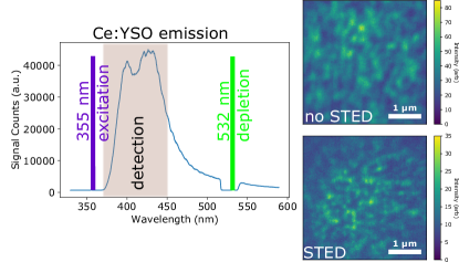
Superresolution Microscopy
Based on similar optical properties of Ce3+:YAG and Ce3+:YSO, superresolution microscopy techniques, such as STED microscopy, which are known to be applicable to Ce3+:YAGKolesov et al. (2018), were used to increase spatial resolution for microscopy of Ce3+:YSO. This was achieved by superimposing a picosecond-pulsed 532 nm doughnut shaped depletion beam on the picosecond-pulsed 355 nm excitation beam. In order to obtain highest increase in optical resolution, laser powers had to be adjusted carefully with respect to each other and the 532 nm laser pulse arrival time had to be delayed with respect to the excitation pulse. The optical detection window for confocal laser scans for both, with and without additional STED beam, stayed unchanged throughout the experiments, and spanned the range between 365 nm-450 nm, as indicated in the left image of Fig. 6. Confocal laser scans depicted on the right side of Fig. 6 show on top the measurement without STED beam and on bottom with STED beam. By comparing individual point spread functions in both scans, we can quantify a resolution enhancement of factor 2. This resolution enhancement, though in principle capable of significantly reducing background noise for our spin spectroscopic studies, was not available in conjunction with spin manipulation. While the high power 532 nm depletion laser was used, no magnetic resonance signals could be acquired. We conjecture, that high power picosecond pulses at 532 nm prevent the optical spin initialization and readout, potentially through ionization of Ce3+ and surrounding traps.
Hahn-Echo Measurement Sequence
For Hahn-Echo measurements, the signal was acquired in a balanced measurement sequence, where the final pulses in the sequence alternatingly projected the spin to or , by applying either or MW pulses at the end of the sequence, depicted in Fig 7b. These two signals are subsequently subtracted from each other, which corresponds to the coherence of the electron spin at a time , defined as the average value of the transverse spin component .

Simulation
In this part, we give the simulation method of the spin decoherence of Ce3+ ion subjected to a YSO nuclear spin bath. The Ce3+ ion in the YSO crystal, substituting one yttrium, forms a defect center. Due to the crystal field, the fine structure of the Ce3+ ion splits further to three doublets. The ground doublet state subspace is our key concern. Its Hamiltonian is
| (1) |
where is the Bohr magneton, is the Planck constant, is the magnetic field, is the lowest doublet state spin operator, and is the effective g-factor of Ce3+ Pidol et al. (2006); Wen et al. (2014)
| (2) |
The Ce3+ ion is surrounded by a dense nuclear spin bath, which consists of nuclear spins and nuclear spins. These nuclear spins interact with Ce3+ ion through magnetic dipole-dipole interactions Siyushev et al. (2014)
| (3) |
where is the magnetic constant, is the gyromagnetic ratio of the -th nuclear spin, is the -th nuclear spin operator, is the distance between the Ce3+ defect and the -th nuclear spin, and is the corresponding unit vector. The nuclear spin bath itself is also governed by Hamiltonian
| (4) |
where is the distance between two nuclear spins, and is the corresponding unit vector.
Now, we investigate the decoherence of Ce3+ under the dynamical decoupling sequences CPMG-, where its decoherence is mainly contributed by its surrounding nuclear spins. In the experiment, we initialize the defect Ce3+ centers to a polarized state, and employ a microwave pulse to prepare the defect Ce3+ to a coherence state. At experimental temperatures the nuclear spins are hardly polarized so that their initial state can be well modeled as a high-temperature mixed state , where is the identity operator for the -th nuclear spin. Driven by the Hamiltonian under CPMG-, the decoherence of the defect centers can then be obtained as Zhao et al. (2012b); Siyushev et al. (2014)
| (5) |
where is the pulse interval of the CPMG- sequences, and , defined by
is the conditional Hamiltonian of projected to the eigenstate of Zhao et al. (2012b); Siyushev et al. (2014). With the help of Cluster-Correlation Expansion (CCE) method Zhao et al. (2012b); Yang and Liu (2009), the decoherence of the Ce3+ is numerically calculated as
| (6) |
where is the decoherence of Ce3+ induced by the nuclear spin cluster . When the concerned time scale is far less than microseconds (corresponding to interaction strength for single nuclear spins), the decoherence of the Ce3+ can be well described by a non-interacting nuclear spin bath. In our system, we can use such a non-interacting approximation because the Zeeman interaction strength and the hyperfine interaction strength between Ce3+ and the nuclear spins is much larger than the interaction within the bath spins.
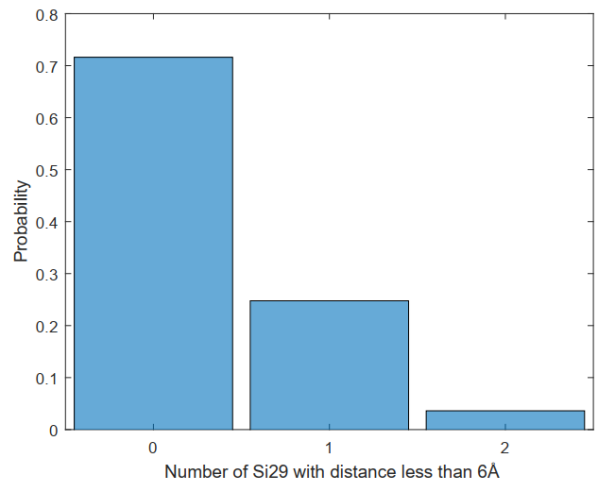
As mentioned in the main text, the cerium density in the sample can lead to more than one Ce3+ ion to be probed within the focal volume. As a consequence, the decoherence signal can in principle result from more than one Ce3+ ion. The different Ce3+ ions in the same spot share the same yttrium nuclear spin environment. However, different Ce3+ ions can experience quite different nuclear spin environments, based on statistcal occurance of . To account for this, for simulations for ”ion B” shown in Fig. (4) in the main text, we assumed there are two Ce3+ ions in a lattice but only one Ce3+ ion has a proximal with distance Å. While for ”ion A”, we assumed there is only one Ce3+ ion and no proximal nearby Ce3+ in a distance smaller than 6 Å. While we cannot distinguish between one or two coupled with distance closer than 6 Å to the Ce3+ ion with the demonstrated spin spectroscopy, it is more likely to sense exactly one single nuclear spin. Based on the 5 % abundance of , we have estimated the likelyhood to detect no at all, exactly one or two , shown in Figure 8.
The coherence difference between two different ions reflects the difference from proximal nuclear spin environment. With increasing CPMG pulse number, we expect the coherence difference to have sharp coherence dips via a direct CCE simulation. However, the experimentally observed coherence dips are much more shallow than the theoretical prediction for CPMG-5. One possible reason is external noise for proximal , for example magnetic field fluctuations introduced by MW manipulation to Ce3+. To take noise into account, we phenomenologically add a relaxation and dephasing mechanism to the dynamics of Lindblad (1976); Breuer and Petruccione (2002); Wang and Yang (2015)
| (7) |
where , are the Pauli matrices of the proximal nuclear spin, and and are the dephasing and relaxiation rates. Our simulation, shown as solid lines in the main text in Fig. 4(b)-(d), uses (which ranges at a similar magnitude in the NV center system Wang and Yang (2015)).
References
- Morton et al. (2008) J. Morton, A. M. Tyryshkin, R. M. Brown, S. Shankar, B. W. Lovett, A. Ardavan, T. Schenkel, E. E. Haller, J. W. Ager, and S. A. Lyon, Nature 455, 1085 (2008).
- Maurer et al. (2012) P. C. Maurer, G. Kucsko, C. Latta, L. Jiang, N. Y. Yao, S. D. Bennett, F. Pastawski, D. Hunger, N. Chisholm, M. Markham, D. Twitchen, J. Cirac, and M. Lukin, Science 336, 1283 (2012).
- Awschalom et al. (2018) D. D. Awschalom, R. Hanson, J. Wrachtrup, and B. B. Zhou, Nat. Photonics 12, 516 (2018).
- Waldherr et al. (2014) G. Waldherr, Y. Wang, S. Zaiser, M. Jamali, T. Schulte-Herbrüggen, H. Abe, T. Ohshima, J. Isoya, J. Du, P. Neumann, and J. Wrachtrup, Nature 506, 204 (2014).
- Dibos et al. (2018) A. M. Dibos, M. Raha, C. M. Phenicie, and J. D. Thompson, Phys. Rev. Lett. 120, 243601 (2018).
- De Riedmatten and Afzelius (2015) H. De Riedmatten and M. Afzelius, in Engineering the Atom-Photon Interaction (Springer, 2015) pp. 241–273.
- Zhong et al. (2015) M. Zhong, M. P. Hedges, R. L. Ahlefeldt, J. G. Bartholomew, S. E. Beavan, S. M. Wittig, J. J. Longdell, and M. J. Sellars, Nature 517, 177 (2015).
- Kolesov et al. (2012) R. Kolesov, K. Xia, R. Reuter, R. Stöhr, A. Zappe, J. Meijer, P. Hemmer, and J. Wrachtrup, Nat. Commun. 3, 1029 (2012).
- Yin et al. (2013) C. Yin, M. Rancic, G. G. de Boo, N. Stavrias, J. C. McCallum, M. J. Sellars, and S. Rogge, Nature 497, 91 (2013).
- Kolesov et al. (2013) R. Kolesov, K. Xia, R. Reuter, M. Jamali, R. Stöhr, T. Inal, P. Siyushev, and J. Wrachtrup, Phys. Rev. Lett. 111, 120502 (2013).
- Zhong et al. (2018) T. Zhong, J. M. Kindem, J. G. Bartholomew, J. Rochman, I. Craiciu, V. Verma, S. W. Nam, F. Marsili, M. D. Shaw, A. D. Beyer, and A. Faraon, Phys. Rev. Lett. 121, 183603 (2018).
- Zhao et al. (2012a) N. Zhao, J. Honert, B. Schmid, M. Klas, J. Isoya, M. Markham, D. Twitchen, F. Jelezko, R.-B. Liu, H. Fedder, and J. Wrachtrup, Nat. Nanotechnol. 7, 657 (2012a).
- Kolkowitz et al. (2012) S. Kolkowitz, Q. P. Unterreithmeier, S. D. Bennett, and M. D. Lukin, Phys. Rev. Lett. 109, 137601 (2012).
- Taminiau et al. (2012) T. H. Taminiau, J. J. T. Wagenaar, T. van der Sar, F. Jelezko, V. V. Dobrovitski, and R. Hanson, Phys. Rev. Lett. 109, 137602 (2012).
- Car et al. (2018) B. Car, L. Veissier, A. Louchet-Chauvet, J.-L. Le Gouët, and T. Chanelière, Phys. Rev. Lett. 120, 197401 (2018).
- Zhong et al. (2019) M. Zhong, R. L. Ahlefeldt, and M. J. Sellars, New J. Phys. 21, 033019 (2019).
- Childress et al. (2006) L. Childress, M. G. Dutt, J. Taylor, A. Zibrov, F. Jelezko, J. Wrachtrup, P. Hemmer, and M. Lukin, Science 314, 281 (2006).
- Nagy et al. (2019) R. Nagy, M. Niethammer, M. Widmann, Y.-C. Chen, P. Udvarhelyi, C. Bonato, J. U. Hassan, R. Karhu, I. G. Ivanov, N. T. Son, et al., Nat. Commun. 10, 1954 (2019).
- Pla et al. (2013) J. J. Pla, K. Y. Tan, J. P. Dehollain, W. H. Lim, J. J. Morton, F. A. Zwanenburg, D. N. Jamieson, A. S. Dzurak, and A. Morello, Nature 496, 334 (2013).
- Pidol et al. (2006) L. Pidol, O. Guillot-Noël, A. Kahn-Harari, B. Viana, D. Pelenc, and D. Gourier, J. Phys. Chem. Solids 67, 643 (2006).
- Drozdowski et al. (2004) W. Drozdowski, A. J. Wojtowicz, D. Wiśniewski, P. Szupryczyński, S. Janus, J.-L. Lefaucheur, and Z. Gou, J. Alloys Compd. 380, 146 (2004).
- (22) , Supplementary Material.
- Yan et al. (2013) Y. Yan, J. Karlsson, L. Rippe, A. Walther, D. Serrano, D. Lindgren, M.-e. Pistol, S. Kröll, P. Goldner, L. Zheng, and J. Xu, Phys. Rev. B 87, 184205 (2013).
- Kolesov et al. (2018) R. Kolesov, S. Lasse, C. Rothfuchs, A. D. Wieck, K. Xia, T. Kornher, and J. Wrachtrup, Phys. Rev. Lett. 120, 033903 (2018).
- Siyushev et al. (2014) P. Siyushev, K. Xia, R. Reuter, M. Jamali, N. Zhao, N. Yang, C. Duan, N. Kukharchyk, A. Wieck, R. Kolesov, and J. Wrachtrup, Nat. Commun. 5, 3895 (2014).
- Schweiger and Jeschke (2001) A. Schweiger and G. Jeschke, in Principles of Pulse Electron Paramagnetic Resonance (Oxford University Press, 2001) pp. 247–255.
- Zhao et al. (2012b) N. Zhao, S.-W. Ho, and R.-B. Liu, Phys. Rev. B 85, 115303 (2012b).
- Kurkin and Chernov (1980) I. Kurkin and K. Chernov, Physica B+C 101, 233 (1980).
- Zhao et al. (2011) N. Zhao, J.-L. Hu, S.-W. Ho, J. T. Wan, and R. Liu, Nat. Nanotechnol. 6, 242 (2011).
- Wang and Yang (2015) P. Wang and W. Yang, New J. Phys. 17, 113041 (2015).
- Ma et al. (2015) W. Ma, F. Shi, K. Xu, P. Wang, X. Xu, X. Rong, C. Ju, C.-K. Duan, N. Zhao, and J. Du, Phys. Rev. A 92, 033418 (2015).
- London et al. (2013) P. London, J. Scheuer, J.-M. Cai, I. Schwarz, A. Retzker, M. B. Plenio, M. Katagiri, T. Teraji, S. Koizumi, J. Isoya, R. Fischer, L. P. McGuinness, B. Naydenov, and F. Jelezko, Phys. Rev. Lett. 111, 067601 (2013).
- Raha et al. (2019) M. Raha, S. Chen, C. M. Phenicie, S. ourari, A. M. Dibos, and J. Thompson, arXiv.org arXiv:1907.09992v1 (2019).
- Wen et al. (2014) J. Wen, C.-K. Duan, L. Ning, Y. Huang, S. Zhan, J. Zhang, and M. Yin, J. Phys. Chem. A 118, 4988 (2014).
- Yang and Liu (2009) W. Yang and R.-B. Liu, Phys. Rev. B 79, 115320 (2009).
- Lindblad (1976) G. Lindblad, Comm. Math. Phys. 48, 119 (1976).
- Breuer and Petruccione (2002) H.-P. Breuer and F. Petruccione, The Theory of Open Quantum Systems (Oxford University Press, 2002).