Dynamic detection of a single bacterium: nonlinear rotation rate shifts of driven magnetic microsphere stages
Abstract
We report on a new technique which was used to detect single Escherichia coli that is based on the changes in the nonlinear rotation of a magnetic microsphere driven by an external magnetic field. The presence of one Escherichia Coli bacterium on the surface of a 2.0 magnetic microsphere caused an easily measurable change in the drag of the system and, therefore, in the nonlinear rotation rate. The straight-forward measurement uses standard microscopy techniques and the observed average shift in the nonlinear rotation rate changed by a factor of 3.8.
Magnetic microspheres and nanoparticles have been used for a variety of medical applications and incorporated into various diagnostic techniques Haukanes and Kvam (1993); Olsvik et al. (1994); Gu et al. (2003). While magnetic particles have proven to be extremely useful, they have been generally utilized in techniques that depend on the translational properties of magnetic particles, such as magnetic separation, giant magneto-resistive (GMR) sensors Rife et al. (2003), and magnetic tunnel junctions (MTJ) sensors Shen et al. (2005); however, it is possible through standard microscopy techniques, to monitor the rotational behavior of single magnetic particles or small chains of them Anker and Kopelman (2003); Biswal and Gast (2004); Lapointe et al. (2005); Korneva et al. (2005); McNaughton et al. (2006a). These small magnetic systems have been utilized to improve immunoassays Petkus et al. (2006), to act as micro-mixers Biswal and Gast (2004), study microrheology Lapointe et al. (2005); Behrend et al. (2005) and even to reduce interfering background in fluorescent spectroscopy measurements Anker and Kopelman (2003). While single bacteria have been detected in fluid using nanoparticles Zhao et al. (2004), a dynamic application that has yet to be developed is the detection of single micro-biological agents using magnetic particles. We report on the first measurement of a single bacterium using a rotating micro-stage.
This new method, being development by the authors, is based on the nonlinear rotation that a magnetic microsphere undergoes when driven by a rotating magnetic field McNaughton et al. (2006a, b). The effect occurs when the viscous torque that arises from rotational drag is comparable to the magnetic torque created by the external driving field. At low external driving frequencies, the magnetic particle rotates continuously and is synchronous with the external field, but at sufficiently high external driving frequencies, the particle becomes asynchronous with the driving field. The external driving frequency, where the magnetic particle goes from linear to nonlinear (synchronous to asynchronous) rotation, is dependent on environmental conditions in addition to the particle properties and is given by McNaughton et al. (2006a, b)
| (1) |
where is the magnetic moment, is the external magnetic field, is the shape factor, is the dynamic viscosity, and is volume. The rotational dynamics of an actively rotated magnetic particle are then given by
| (4) |
where is the particle’s average rotation rate and is the driving frequency of an external magnetic field. Equation 4 holds for low Reynolds number environments () and for our system .
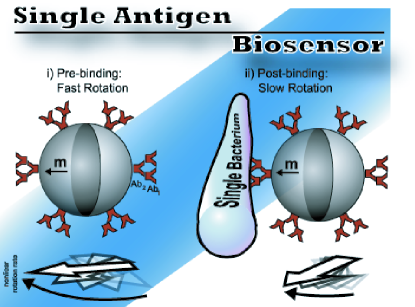
Nonlinear rotation occurs when Shelton et al. (2005) and we propose that this rate can be used to detect single microbiological agents. The parameters that are important in biological detection are shape and volume because of the drag changes that occur when a bacteria binds to a microsphere. So, when a bacteria attaches to a nonlinear rotating magnetic microsphere, the micro-stage’s volume and shape are drastically changed, which produces more drag and, therefore, the rotation rate slows considerably. This is shown schematically in Figure 1. The technique is dynamic in the sense that a change in drag causes a direct change in the nonlinear rotation rate. Past measurements have shown that this technique can measure a change of drag caused by an attachment of a 1.0 particle to a 1.9 nonlinear rotating magnetic microsphere McNaughton et al. (2006c).
The ability to develop sensitive diagnostic techniques using dynamic systems has been a topic of high interest. One related technology that has demonstrated extreme sensitivity in air and vacuum environments is nanoelectromechanical systems (NEMS) Ekinci and Roukes (2005). NEMS have been used to measure the mass of single micro-biological agents like antibodies, viruses, and bacteria Ilic et al. (2000, 2001, 2004). One of the NEMS detection schemes utilize the fact that the resonant oscillations change when a micro-biological agent attaches, but the sensitivity of such devices are drastically affected when operated in fluidic environments Vignola et al. (2006). The idea underlying nonlinear rotating magnetic particles can be used in a similar way, namely when a biological agent attaches to the magnetic particle, the nonlinear rotation frequency changes, but in such systems sensitivity is unaffected, rather helped by drag. This allows for single biological agent detection in fluidic environments. Thus, in this letter we show that the nonlinear rotation frequency of single magnetic microspheres on average rotates significantly slower when a single bacterium is attached to their surface.
A 20 aliquot of 2.0 ferromagnetic microspheres functionalized with goat antimouse IgG (Spherotech IL) was spread onto a precut microscope slide and coated with 50 of Al. The sample was placed in a uniform magnetic field of 1.4 so that the magnetization would be perpendicular to the microscope glass. The spheres were then rinsed with phosphate buffer solution (PBS) at a pH of 7.2 and suspended in 500 of PBS. The suspended sample was centrifuged at 9000 for 8 and resuspended in 500 of PBS at a pH of 7.2. The sample was centrifuged once more at 9000 for 8 and the supernatant was removed. 100 of mouse anti-E. Coli IgG (Cortex Biochem, San Diego, CA) was added to the pellet of magnetic microspheres. The primary antibody and the magnetic microspheres were allowed to incubate at room temperature for 4 . The excess primary antibody was removed by centrifuging the sample at 9000 for 8 and the supernatant was discarded. Finally, the magnetic microspheres were resuspended into 500 of PBS. At each of the above stages the sample was vortexed at 3000 for 15 seconds.
To make the bacteria fluorescent, a DsRed plasmid was used with Escherichia Coli BL21(DE3) following previously described transformation procedures Shaner et al. (2004). The bacteria were allowed to reproduce until the sample had an optical density of .67 at 600 nm and was stored at 4 . The magnetic microspheres with the anti-E. Coli antibody were mixed 1:1 with the now fluorescent E. Coli described. To make binding more probable, the sample was centrifuged at 9000 for 8 . The sample was then vortexed at 3000 for 15 and allowed to incubate. The resulting sample had many single microspheres with 1-5 E. Coli bound to their surface, where visual analysis was used to confirm the presence of a single bacterium.
Two homemade 100 thick fluidic cells were fabricated: one fluidic cell contained the magnetic microsphere solution before bacteria was added and the other had magnetic microspheres with bacteria bound to their surfaces. Before being placed in the fluidic cells, the samples were mixed with glycerol so that the glycerol-water mass fraction was 0.5. The nonlinear rotation rates for 20 single magnetic microspheres, without bacteria, were obtained by monitoring the intensity fluctuations caused by light reflecting off of the aluminum half-shell. From the other fluidic cell, 20 nonlinear rotation rates were obtained for single magnetic microspheres with one E. Coli bound to their surfaces by monitoring the intensity fluctuations caused by the bacteria fluorescence (for more experimental and data analysis details see Refs Anker and Kopelman (2003); McNaughton et al. (2006a, b)). The rotation rates were then averaged and compared to determine the rotation rate changes caused by the bacteria.
The theory for a single magnetic particle rotating in response to an external driving field is well developed McNaughton et al. (2006a, b); Cēbers and Ozols (2006), but measurements have not previously been made for the case of a magnetic particle attached to a bacteria. Figure 2(a) shows the average rotation rate of such a system for increasing external driving frequencies. The data is in good agreement with the fit determined from Equation 4 and the critical slipping rate, , was found to be 1.27 . This measurement shows that when a bacteria is bound to the surface of a magnetic microsphere, the system can still be analyzed using previously developed theory. Thus, a change in rotation rate can be used to detect bacteria.
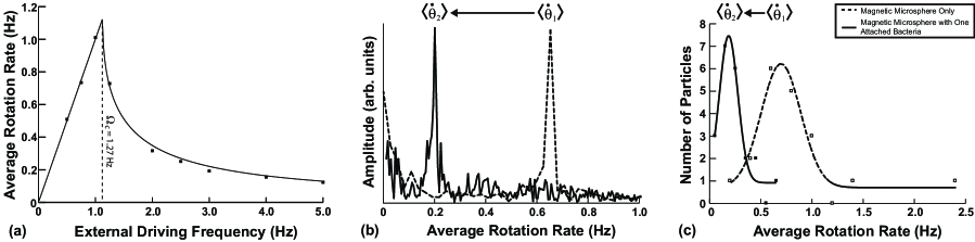
While the entire range of frequencies for magnetic particles with and without bacteria could be scanned as was done in Figure 2(a), it is much faster and more straight-forward to only measure the value of the nonlinear rotation rate, , at a given external driving frequency of . Figure 2(b) shows this measurement for a typical magnetic microsphere with and without a single bacteria attached to its surface. Figure 2(c) shows the curves for the rotation rate of 20 particles in a fluidic cell with bacteria and for 20 particles in one without bacteria. The presence of the bacteria on the surface of the magnetic microspheres caused a measurable change in the average rotation rate, namely the average rate of the particles at a driving frequency of 4.0 changed from to , a factor of 3.8. This change in rotation rate is similar in value to our previous measurements on a 1.0 particle that was attached to a single 1.9 ferromagnetic microsphere McNaughton et al. (2006c). Once a bacteria is attached to a magnetic microsphere, this technique could also be used to monitor single bacteria growth, which could have significant application for the study of single bacteria growth dynamics and in antibiotic susceptibility measurements.
The ability to use the change in nonlinear rotation of magnetic particles to detect bacteria has been demonstrated. The nonlinear rotation rate of 2.0 magnetic microspheres changed on average from 0.72 without a bacterium to 0.19 Hz with a single bacterium attached, where the driving oscillatory magnetic field was at a frequency of 4.0 .
The authors would like to thank Carol A. Fierke, Marcy Hernick, and Tamiika K. Hurst for help with the bacteria growth and transformations. Funding was provided by NSF-DMR 0455330. Related information, such as videos will be available online at http://www.umich.edu/koplab/moons.htm.
References
- Haukanes and Kvam (1993) B. Haukanes and C. Kvam, Bio-Technology 11, 60 (1993).
- Olsvik et al. (1994) O. Olsvik, T. Popovic, E. Skjerve, K. Cudjoe, E. Hornes, J. Ugelstad, and M. Uhlen, Clinical Microbiology Reviews 7, 43 (1994).
- Gu et al. (2003) H. Gu, P. Ho, K. Tsang, C. Yu, and B. Xu, Chemical Communications 15, 1966 (2003).
- Rife et al. (2003) J. Rife, M. Miller, P. Sheehan, C. Tamanaha, M. Tondra, and L. Whitman, Sensors and Actuators A 107, 209 (2003).
- Shen et al. (2005) W. Shen, X. Liu, D. Mazumdar, and G. Xiao, Applied Physics Letters 86, 253901 (2005).
- Anker and Kopelman (2003) J. Anker and R. Kopelman, Applied Physics Letters 82, 1102 (2003).
- Biswal and Gast (2004) S. Biswal and A. Gast, Anal. Chem 76, 6448 (2004).
- Lapointe et al. (2005) C. Lapointe, N. Cappallo, D. Reich, and R. Leheny, Journal of Applied Physics 97, 10 (2005).
- Korneva et al. (2005) G. Korneva, H. Ye, Y. Gogotsi, D. Halverson, G. Friedman, J. Bradley, and K. Kornev, Nano Lett 5, 879 (2005).
- McNaughton et al. (2006a) B. McNaughton, K. Kehbein, J. Anker, and R. Kopelman, Journal of Physical Chemistry B 110, 18958 (2006a).
- Petkus et al. (2006) M. Petkus, M. McLauchlin, A. Vuppu, L. Rios, A. Garcia, and M. Hayes, Anal. Chem 78, 1405 (2006).
- Behrend et al. (2005) C. Behrend, J. Anker, B. McNaughton, and R. Kopelman, Journal of Magnetism and Magnetic Materials 293, 663 (2005).
- Zhao et al. (2004) X. Zhao, L. Hilliard, S. Mechery, Y. Wang, R. Bagwe, S. Jin, and W. Tan, Proceedings of the National Academy of Sciences 101, 15027 (2004).
- McNaughton et al. (2006b) B. McNaughton, R. Agayan, J. Wang, and R. Kopelman, Sensors and Actuators B, In Press (2006b).
- Shelton et al. (2005) W. Shelton, K. Bonin, and T. Walker, Physical Review E 71, 36204 (2005).
- McNaughton et al. (2006c) B. McNaughton, R. Agayan, V. Stoica, R. Clarke, and R. Kopelman, In Preparation (2006c).
- Ekinci and Roukes (2005) K. Ekinci and M. Roukes, Review of Scientific Instruments 76, 61101 (2005).
- Ilic et al. (2000) B. Ilic, D. Czaplewski, H. Craighead, P. Neuzil, C. Campagnolo, and C. Batt, Applied Physics Letters 77, 450 (2000).
- Ilic et al. (2001) B. Ilic, D. Czaplewski, M. Zalalutdinov, H. Craighead, P. Neuzil, C. Campagnolo, and C. Batt, Journal of Vacuum Science & Technology B: Microelectronics and Nanometer Structures 19, 2825 (2001).
- Ilic et al. (2004) B. Ilic, Y. Yang, and H. Craighead, Applied Physics Letters 85, 2604 (2004).
- Vignola et al. (2006) J. Vignola, J. Judge, J. Jarzynski, M. Zalalutdinov, B. Houston, and J. Baldwin, Applied Physics Letters 88, 41921 (2006).
- Shaner et al. (2004) N. Shaner, R. Campbell, P. Steinbach, B. Giepmans, A. Palmer, and R. Tsien, Nature Biotechnology 22, 1567 (2004).
- Cēbers and Ozols (2006) A. Cēbers and M. Ozols, Physical Review E 73, 21505 (2006).