Dynamic re-wiring of protein interaction:
The case of transactivation
Gabriele Scheler
ISLE
Ventura Hall 25
200 Panama Street
Stanford, Ca. 94305
Abstract
We are looking at local protein interaction networks from the perspective of directed, labeled graphs with quantitative values for monotonic changes in concentrations. These systems can be used to perform stability analysis for a stable attractor, given initial values. They can also show re-configuration of whole system states by dynamic insertion of links, given specific patterns of input. The latter issue seems particularly relevant for the concept of multistability in cellular memory. We attempt to show that this level of analysis is well-suited for a number of relevant biological subsystems, such as transactivation in cardiac myocytes or G-protein coupling to adrenergic receptors. In particular, we analyse the ’motif’ of an ”overflow gate” as a concentration-dependent system reconfiguration.
1 Introduction
We are interested in cases, where the intracellular pathways established by protein interaction and small messenger molecules show a dynamic change (’re-wiring’) due to external conditions. For two distinct major pathways, the G-protein coupled pathway activated by ligand-bound receptor molecules and the MAP kinase pathway activated by tyrosine kinases, it has been shown that regulatory interactions arise under certain conditions, in a process known as ’transactivation’([4, 12]). Transactivation is thus an ideal model case to study dynamic re-wiring of protein interactions, its conditions, and its functional consequences. Another related process, the antagonism of G-protein coupled receptors, which occurs as a submodule in transactivation, provides another opportunity to analyse the dynamics of protein interaction.
The computational analysis of protein interaction has focused on the establishment of protein interaction networks and their analysis by search for local motifs or subgraphs in attempts to link specific subgraphs to functional modules [9]. Showing how and why functional subgraphs change will add a much needed dynamical perspective to the analysis of protein interaction networks. To analyse these patterns seems a useful step in order to cut down on the complexity of the temporal dynamics of intracellular signaling in a given situation.
In many ways, intracellular networks need to function as adaptive control systems: keeping key parameters within tightly controlled bounds in response to many extracellular events. However, to provide memory, they also need to be multistable, i.e. function within different regimes. Probabilistic re-wiring of subgraphs provides an adequate representation for the dynamics of interactions to allow to analyse changing control pathways and their stability (cf. [11]).
2 Transactivation of the MAP kinase pathway by G proteins: A cascade of overflow gates
Tyrosine kinases (such as EGFR) occur in membrane-bound position where they can be activated by both extracellular and intracellular events. They comprise an evolutionarily conserved, ’old’ type of receptor with little specificity in conditions for activation (they are typically activated by up to six different ligands) and effects on an important intracellular pathway, the MAP kinase pathway, which is involved in cellular housekeeping functions, such as initiating growth or apoptosis.
The large protein family of G-protein coupled receptors (GPCRs) consists of membrane-bound proteins which change their conformational state by being bound with specific ligands in extracellular concentrations and effecting the separation of oligomeric G proteins (, and proteins) into and components [10]. There are usually at least 2, up to 7 or so different GPCRs that bind to the same extracellular ligand with considerable specificity. The different G proteins they interact with have, among other effects, antagonistic functions on downstream signaling: proteins augment the adenylyl cyclase, cyclic AMP and protein kinase A pathway and proteins suppress this pathway.
We propose that the basic function of this GPCR antagonism is a regulation of parametric ranges for the common substrate in downstream signaling, the cAMP/PKA pathway(’GPCP’). It is remarkable that for each ligand, there exists usually both an ’excitatory’, -coupled receptor, and an ’inhibitory’ coupled receptor [10]. Mostly, the Gi-coupled receptor has a lower affinity to the ligand, and thus becomes activated only at higher concentrations. Thus there is an overflow-dependent activation of an additional protein interaction, in this case with a suppressive effect on a common outcome with a pre-existing interaction (see Fig. 1A).
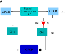
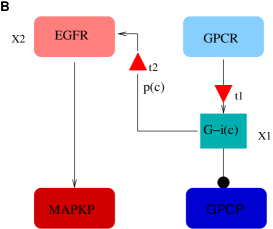
The basic principle in ’transactivation’ relies on a variation of the same concept: A high level of protein activity leads to the insertion of an interactive link from proteins to the EGF receptor, resulting in an additional, augmenting activation of this receptor [4, 12]. Functionally, this corresponds to the insertion of a regulatory interaction between the GPC and the MAPK pathways triggered by the concentration of a protein in one of the pathways (see Fig. 1B,t2).
We want to establish this basic mechanism of a concentration-activated protein interaction in very general terms, as a simple, conserved motif in the dynamics of protein interaction:
-
1.
A link between and becomes established with probability , where depends in a functional manner on a concentration related to . (concentration-dependent gate, overflow gate)
The concentration may be identical to a local concentration of as in Fig. 1B or the concentration of an extracellular ligand (or small molecule, such as cAMP or calcium) that determines the activation state (conformational change) of (as in Fig. 1A). The probability p will in many cases be determined by a sigmoidal function, which establishes a lower threshold for the probability to increase and an upper threshold (saturation) for it to remain constant (U-shaped or other functions are also possible). A local protein interaction network may operate preferentially in a low regime - where the sigmoidal function operates mainly as a threshold - or in a higher regime - where the sigmoidal function operates mainly as a linear dependence.
There are potentially other motifs, which require only a probabilistic interpretation of concentrations: e.g. a pool concentration may interact with two different partners according to a fixed partitioning (, ), with the possibility of one of these connections acting as an informative control signal, ’telling’ another pathway about the level of activation of another pathway (Example: [1, 8, 7]).
Note however that the choice of a definition involving probabilities in determining protein interaction does not capture the time course of interactions explicitly (see below, section 3 for a discussion).
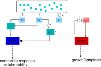
Transactivation occurs in many types of cells [12], and involves a number of additional processes [12, 15]. Within the context of cardiac myocytes, another prominent process is aptly categorized as an ’overflow gate’: The native ligand, noradrenaline, activates both - (-coupled) and -(-coupled) adrenergic receptors, which are antagonistically related. In addition, there are two different variants of -adrenergic receptors, -1 and -2, where -2 additionally activates proteins, when being continually or highly activated [16]. In cardiac myocytes, different cellular outcomes (increased contractile responses [3], growth and apoptosis [13, 5]) are associated with the level of activation of the MAPK pathway system. We see, interestingly, that the overactivation associated with a harmful response is being protected by a system consisting of a three-degree cascade of overflow gates (see Fig. 2A).
3 Temporal dynamics of an ’overflow gate’
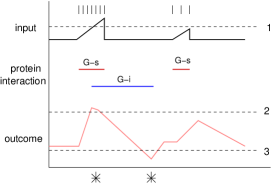
By focusing on the probability of a protein interaction, we lose the temporal fine structure of concentration changes in establishing a link that can be derived from kinetic reaction rate models or similar dynamical equations. For instance, not only are G-s coupled receptors activated by lower levels of ligands, they also desensitize more quickly, and undergo faster recycling at the membrane. G-i coupled receptors, which require higher levels of ligands, desensitize more slowly, and have slower turnover rates as well. The fine temporal structure of this process is depicted schematically in Fig. 2B. Initial G-s activation is counteracted after a delay by G-i activation if the signal keeps increasing. G-i activation lasts for a considerable longer period of time which may result in hysteresis of the response. Instead of G-s and G-i interacting to keep a stable range of outcome no matter how strong the signal, we now have time courses for the outcome where high values may be tolerated, if they are counteracted in time by an antagonistic force. We can see that the requirements of stable control parameters can be temporarily lifted to create significant high or low signals. The signal also has a characteristic time course with a rapid increase and a slow dynamics of suppression (common in a system of antagonistic regulation). Other patterns for time courses are, for instance, slow rise times, which correspond to temporal buffers. Thus the fine temporal structure can also be analysed with the goal of discovering most simple motifs with considerable generality. Biochemically, protein interactions can be adequately modelled and simulated by reaction-rate equations, such as the Michaelis-Menten formulation. Integrating explicit rate information into a probabilistic dynamic structure also bridges the gap to an actual simulation model of protein interaction.
4 Summary
We have started from a well-described model system (transactivation in cardiac myocytes) in an attempt to find simple, general dynamic motifs, beyond the paradigm case of the ’feedback loop’. We have also outlined how this information can be included in existing protein interaction networks (e.g. [2]), using a probabilistic formulation for concentration-dependent dynamic re-wirings, and specifically analysed the motif of an ’overflow switch’ with three examples.
Actual protein interaction occurs in tight local structures (using scaffolds and other properties of the actin cytoskeleton) which often give the impression of highly ordered, well-designed machinery. This is particularly true for membrane-bound and membrane-close proteins (in contrast to cytosolic proteins which diffuse more freely). Protein networks only provide a static picture of possible interactions. A dynamic view, however, can be achieved on different levels. One goal is to describe control interactions and adaptive stability or multistability within the system. We have tried to show how to identify and define dynamic motifs which do two things: they insert a link in a subgraph and they specify the conditions for the link to become active. For this we used quantities(local concentrations) and (sigmoidal or other) probabilities of interaction. This allows the specification of memory in the system, and goes beyond a static description of all possible interactions, and their characteristics.
To fully reconstruct a complex biological function such as neural adaptivity, we will also need to look at the time-courses of changes and integrate over different time-scales. Thus, another goal of a dynamic network is to provide an integration with simulation models on the level of kinetic rate equations. Here again, we may look for conserved motifs of characteristic time curves and their associated function.
Engineering and evolution share an approach of building complex systems from repetition of simple modules [14], [6]. The real challenge lies in understanding the integrative principles in cellular systems, which may be significantly different from the planned design approach characteristic of engineered systems.
References
- [1] A. J. Barron, S. G. Finn, and S. J. Fuller. Chronic activation of extracellular-signal-regulated protein kinases by phenylephrine is required to elicit a hypertrophic response in cardiac myocytes. Biochem J, 371(Pt1):71–9, Apr 2003.
- [2] C.-S. Chin and M. P. Samanta. Global snapshot of a protein interaction network—a percolation based approach. Bioinformatics, 19(18):2413–2419, 2003.
- [3] C. Communal, W. S. Colucci, and K. Singh. p38 mitogen-activated protein kinase pathway protects adult rat ventricular myocytes against beta-adrenergic receptor-stimulated apoptosis. Evidence for Gi-dependent activation. J Biol Chem, 275(25):19395–400, Jun 2000.
- [4] A. Gschwind, E. Zwick, N. Prenzel, M. Leserer, and A. Ullrich. Cell communication networks: epidermal growth factor receptor transactivation as the paradigm for interreceptor signal transmission. oncogene, 20(13):1594–1600, Mar 2001.
- [5] M. Henaff, S. V. Hatem, and J. J. Mercardier. Low catecholamine concentrations protect adult rat ventricular myocytes against apoptosis through cAMP-dependent extracellular signal-regulated kinase activation. Mol Pharmacol, 58(6):1546–53, Dec 2000.
- [6] J. D. Jordan, E. M. Landau, and R. Iyengar. Signaling Networks: The Origins of Cellular Multitasking. Cell, 103:193–200, 2000.
- [7] V. Karoor, S. F. Vatner, G. Takagi, G. Yang, J. Thaisz, J. Sadoshima, and D. E. Vatner. Propranolol prevents enhanced stress signaling in Gsalpha cardiomyopathy: potential mechanism for beta-blockade in heart failure. J Mol Cell Cardiol, 36(2):305–12, Feb 2004.
- [8] K. H. Lee, N. Lee, S. Lim, H. Jung, Y. G. Ko, H. Y. Park, Y. Jang, H. Lee, and K. C. Hwang. Calreticulin inhibits the MEK1,2-ERK1,2 pathway in alpha 1-adrenergic receptor/Gh-stimulated hypertrophy of neonatal rat cardiomyocytes. J Steroid Biochem Mol Biol, 84(1):101–7, Jan 2003.
- [9] R. Milo, S. Shen-Orr, S. Itzkovitz, N. Kashtan, D. Chklovskii, and U. Alon. Network motifs: Simple building blocks of complex networks. Science, 298:824–827, October 2002.
- [10] A. M. Preininger and H. E. Hamm. G Protein Signaling: Insights from New Structures. Science stke, 2004.
- [11] G. Scheler and J. Schumann. Mechanisms for antagonistic regulation of ampa and nmda-d1 receptor complexes at postsynaptic sites. Proceedings of RECOMB, 2004.
- [12] B. H. Shah and K. J. Catt. GPCR-mediated transactivation of RTKs in the CNS: mechanisms and consequences. Trends in Neuroscience, 27(1):48–53, 2004.
- [13] K. Singh, L. Xiao, A. Remondino, D. B. Sawyer, and W. S. Colucci. Adrenergic regulation of cardiac myocyte apoptosis. J Cell Physiol, 189:257–65, 2001.
- [14] V. Spirin and L. A. Mirny. Protein complexes and functional modules in molecular networks. PNAS, 100(21):12123–12128, 2003.
- [15] P. H. Sugden. Signalling pathways in cardiac myocyte hypertrophy. Ann Med, 33(9):611–22, Dec 2001.
- [16] A. M. Zamah, M. Delahunty, L. M. Luttrell, and R. J. Lefkowitz. Protein kinase A-mediated phosphorylation of the beta 2-adrenergic receptor regulates its coupling to Gs and Gi. Demonstration in a reconstituted system. J Biol Chem, 277(34):31249–56, Jun 2002.