Evidence of defect-induced ferromagnetism in ZnFe2O4 thin films.
Abstract
X-ray absorption near-edge and grazing incidence X-ray fluorescence spectroscopy are employed to investigate the electronic structure of ZnFe2O4 thin films. The spectroscopy techniques are used to determine the non-equilibrium cation site occupancy as a function of depth and oxygen pressure during deposition and its effects on the magnetic properties. It is found that low deposition pressures below 10-3 mbar cause iron superoccupation of tetrahedral sites without Zn2+ inversion, resulting in an ordered magnetic phase with high room temperature magnetic moment.
pacs:
61.05.cj, 75.25.-j, 75.47.-m, 75.50.BbI Introduction
Among the functional materials that are being intensely studied to fabricate novel spintronics devices, the spinel oxides appear as an important alternative to perovskites. Particularly, ZnFe2O4 can be a half-metallic, transparent conductor or ferrimagnetic insulator Yanase and Siratori (1984); L ders et al. (2006). At low dimensions, changing the growth conditions the magnetic and electronic properties of the ferrites can be tuned to enhance the magnetic moment considerably and to produce conducting or insulating materials Ayyappan et al. (2010). These properties are very important for the design and fabrication of multilayer structures to produce spin-polarized currents.
It is well known that bulk ZnFe2O4 crystallizes in the normal spinel lattice and that it is antiferromagnetic with a Néel temperature of about 10 K. Zn2+ occupies tetrahedral (A) and Fe3+ octahedral (B) sites. The interaction between magnetic Fe3+ moments is of superexchange type mediated by O2- ions and results in the
magnetic moments being antiparallel to each other. Since the superexchange interaction is stronger for the more closely situated cations and for the bond angle closer to 1800, the superexchange interaction of A-O-B (JAB) is the strongest, the B-O-B (JBB) is weaker, and that of A-O-A (JAA) is the weakest. Accordingly, the magnetic structure and properties of spinel-type oxides depend strongly on the relative strength of the various superexchange interactions. In a normal spinel, magnetic Fe3+ ions occupy only B-sites and the weak negative superexchange interaction among Fe3+ ions (JBB) dominates the magnetic properties of ZnFe2O4 leading to a low Néel temperature and consequently, paramagnetic behavior at room temperature.
The situation is different, however, when normal spinel
ferrite becomes nanosized. Net magnetization at room temperature
can be obtained for nanoparticles obtained by different methods
such as, mechanical synthesis
Yao et al. (2007); Stewart et al. (2007.); Shim et al. (2006), solvothermal
method Blanco-Gutirrez
et al. (2011), solgelShim et al. (2006),
co-precipitation Kamiyama et al. (1992); Ayyappan et al. (2010) and thin
films deposited by sputtering Nakashima et al. (2007) and by pulsed
laser deposition Chen et al. (2008). Several works have suggested
that, in this case, the ferrite displays a nonequilibrium
cation distribution among the tetrahedral and octahedral sites
altering its long-range magnetic ordering. As a consequence the
magnetic response is drastically enhanced. It was reported that
when the particle size decreases Fe3+ occupancy of both A and
B sites in the nanocrystalline state changes and Zn ions are
transferred from their equilibrium position sites A to B sites.
However, there is still some lack of clarity concerning Zn
non-equilibrium positions and their magnetic effects. It is
thought that the occupation of B-sites by Zn2+ brings
Fe3+ ions in both A- and B-sites, and the strong
superexchange interaction between Fe3+ ions in A- and
B-sites forces Fe in B sites to align ferromagnetically causing
high magnetization at room temperature Lotgering (1966); Ligenza (1976); Kamiyama et al. (1992); Shim et al. (2006); Stewart et al. (2007.). In
addition, several authors reported the clear dependence of
magnetic response on the preparation process, apart from the
particle size Kundu et al. (2003); Shenoya et al. (2004). Furthermore, it
was found that both, the magnetic properties and cation
distribution are extremely dependent on deposition conditions
Chen et al. (2008); Takaobushi et al. (2006).
Despite progress in the characterization, the spin configuration
of nanosized ferrites is still unclear and under study
Kamazawa et al. (2003). As the spinel oxides are ferrimagnets
involving two different sublattices, the resulting magnetic moment
is strongly perturbed by defects in the structural lattice. In
addition, there has been increasing evidence that magnetic order
can be triggered by certain defects in materials that are
nominally non magnetic like certain oxides Fernandes et al. (2009); Khalid et al. (2009); Ramos et al. (2010) or pure graphite Ohldag et al. (2010).
In a thin film deposition process many of these defects can be
generated. Particularly, it was recently suggested that oxygen
vacancies affect the magnetic properties of synthesized Zn-ferrite
films grown at various oxygen pressures Sultan and Singh (2009). Also
the oxygen vacancies could be responsible for the semiconducting
behavior found in ZnFe2O4 thin films with high
saturation magnetization at room temperature and high Curie
temperature Chen et al. (2008). There is clearly some need of
employing new experimental procedures using different
techniques that can provide complementary information that
may clarify several open issues in the subject Darko Makovec et al. (2010).
X-ray absorption near-edge structure (XANES) technique together
with simulations based on ab-initio XANES calculations such as
code FEFF8.2 are powerful tools to identify the modification
of Zn2+ and Fe3+ distribution from the equilibrium to
the non-equilibrium
state Stewart et al. (2007.); Nakashima et al. (2007); Figueroa and Stewart (2009); Akhatar et al. (2009). Additionally, Grazing Incidence X-ray Fluorescence (GIXRF) yield the composition, thickness and density of thin films Als-Nielsen et al. (1994); Stoev and Sakurai (1999).
XANES combined with GIXRF results in an interesting method
to relate thickness-dependent electronic structure with magnetic
properties Souza-Neto et al. (2009). XANES is not a surface technique
by itself, since the attenuation length of hard X-rays is a few
micrometres in any material. However, in the grazing-incidence
geometry near the critical angle for total reflection, the X-ray
beam is confined within a few nanometres of the surface. For film
studies, this confinement has the considerable advantage of
minimizing the substrate contribution.
In this work, the structural and magnetic properties of ZnFe2O4 films fabricated by pulsed laser deposition using different oxygen partial pressures are reported and discussed. XANES spectroscopy combined with GIXRF was used to determine the non-equilibrium cation site occupancy as a function of depth and its effects on the magnetic properties. Our results show that the presence of defects as oxygen vacancies or iron situated on normally non-occupied A sites in the spinel structure could be the cause for the magnetism in the films.
II Experimental
The ZnFe2O4 films were fabricated by
pulsed laser deposition (PLD) from a stoichiometric target onto MgO (001) substrates. Substrate temperature was 5000C and the oxygen partial pressure was varied between 10-5 and 10-1
mbar. After deposition the samples were cooled to room temperature in
vacuum at a pressure of about 10-7 mbar.
X-ray diffraction measurements employing Cu Kα radiation in a Philips X-Pert were made for structural characterization
difractometer. Scanning Electron Microscopy, Supra 55VP, with Energy Dispersive Spectroscopy (EDS) was employed to study the surface morphology and spectral analysis. Magnetization measurements were made in a Quantum Design model MPMS-7 SQUID magnetometer. Magnetic
fields were applied parallel to the films.
Electrical resistance measurements were performed using a Keithley 2182A Nanovoltmeter and a
Keithley 6221 current source. Electrical contacts were
made with gold wires clenched with indium. We have measured the
resistance with a two-probe techniques.
GIXRF measurements were carried out at the XRF Fluorescence beamline of the LNLS
(Campinas, Brazil), using a monochromatic X-ray beam of 9.7 keV. The
setup includes 150 m-vertical and 4 mm slits limiting the beam size
on the sample mounted on a high precision goniometer. Angular scans
around the critical angle of total (external) reflection were
performed (between 0 and 2 degrees). The fluorescence emissions were
collected using a 15 element Ge detector. After the angular scan,
XANES Fe K edge (7112 eV) and Zn K edge (9659 eV) spectra in
fluorescence mode were collected at different grazing angles using a
Si (111) channel-cut monochromator. The incident beam intensity and
the energy calibration were monitored using an ion-chamber and a Co (or
Zn) metal standard. The reflected beam and the Co (or Zn) fluorescence
emission were collected using a second ion-chamber and a 15 element Ge
detector, respectively.
Room temperature EXAFS (Extended X-ray Absorption Fine Structure) spectra at the
Zn K-edge were recorded in flourescence mode using a Si(111) monochromator at the XAS2 beamline of the
LNLS (Laboratorio Nacional de Luz Sincrotron) in Campinas, Brazil. In order to estimate the X-ray penetration depth
for each incidence angle, the attenuation length as a function of incidence angle was estimated using the X-ray database
of Lawrence Berkeley National Laboratory Henke et al. (1993).
III Results and discussion
X-ray diffractometry indicated epitaxial growth without any traces of secondary phases. -scans of the ZnFe2O4 (511)
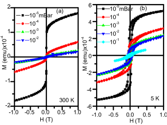
refection showed a fourfold symmetry of the film. Figure 1 shows
the magnetic moment of the films as a function of applied field at
room temperature (a) and at 5 K (b). The diamagnetic signal
from the substrate was subtracted. The S-shaped M-H curves are evidence for ferrimagnetic order. In addition, a linear high-field response
in room temperature measurements indicated the presence of a paramagnetic component. This was also deduced from vs. measurements
(not shown here). The magnetic response diminishes by increasing the oxygen partial pressure from 10-5 to 10-1 mbar as seen by the
decreasing of the magnetic moment by more than one order of magnitude. While at room temperature the coercivity is small,
at low temperatures the films are magnetically rather hard with coercive fields between 447 mT and 1000 mT when deposition pressure varied from P = 10-5 to 10-1 mBar . The drastic reduction of coercive field at room temperature might indicate the existence of magnetic clusters and a blocking mechanism such as has been observed in references [Shim et al., 2006] and [Nakashima et al., 2007] for similar systems.
All films are insulating with room temperature resistivity larger than = 100 m. Sample ZF02 grown under 10-4 O2pressure, had
the smallest electrical resistance and we could measure its behavior under applied magnetic fields between 0.2 and 0.7 T and under light irradiation
between 2.8 and 3.5 eV. The sample did neither present magnetoresistance nor photoconductivity in the range of fields and energies applied.
SEM micrographs show uniform surfaces in all samples. These results suggest that the sample has a spatial uniform magnetic phase at room temperature and that the energy gap in this sample is larger than 3.5 eV.
In table I we summarized the results on characterization of samples labeled as ZFO1 to ZFO5.
| sample | p | t | Fe/Zn | M(RT)10-4 | M(5 K)10-4 | |
|---|---|---|---|---|---|---|
| (mbar) | (nm) | (emu) | (emu) | |||
| ZFO1 | 10-5 | 57 | 1.8 | 1.4 | 5.0 | |
| ZFO2 | 10-4 | 51 | 2.3 | 0.2 | 2.5 | |
| ZFO3 | 10-3 | 43 | 1.9 | 0.1 | 1.9 | |
| ZFO4 | 10-2 | 36 | – | – | 1.5 | |
| ZFO5 | 10-1 | 17 | 1.8 | – | 0.3 |
Using the thicknesses in table I and the magnetic measurements, the estimated magnetization
for sample ZFO1 are 100 emu/cm3 and 360 emu/cm3 at room temperature and 5 K respectively. These are large values considering that ZnFe2O4 with normal spinel structure is an antiferromagnet.
Figure 2 shows the intensity of Fe and Zn lines as a
function of incident angle for all films. The intensities are in
arbitrary units and there is not direct relationship between
intensities and concentration. The angle where a jump
in intensity was observed (approximately 0.32 degree) corresponds to the total reflection
condition. It can be observed that Zn/Fe fraction remains constant for all angles indicating a uniform distribution of both
cations in depth. Furthermore, a Zn enrichment with the increase of oxygen pressure can be seen: the curve
corresponding to Zn almost coincides with the Fe one for 10-5mbar
and this is above the Fe one for P=10-1 mbar. The XRF results were confirmed by EDS (see table I), although it can be seen that the ratio Fe to Zn is close to 2:1 independent of the O2 growth conditions.
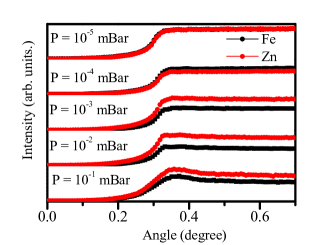
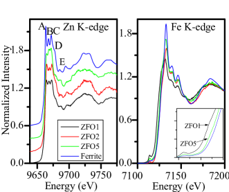
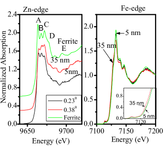
Figure 3 exhibits Zn (right) and Fe (left) K-edge XANES spectra, corresponding to samples 1, 2 and 5 taken at an incidence angle above total reflection (0.38 degree). The attenuation length for this angle was estimated as 35 nm, and then these spectra contain information from the inner of the film. The spectrum of powder ZnFe2O4 normal spinel was included for comparison. Zn K-edge spectra of normal spinel have three resolved peaks, A, B and C (indicated in Fig. 1) at around 9664, 9668 and 9672 eV, respectively, a shoulder at around 9677 eV (peak D), plus additional structure at higher energies (peak E). Zn K-edge spectra of samples 1 and 2 have similar characteristics of normal spinel. This indicates that in these films, Zn ions are mostly on A sites. In the case of sample ZFO5 the increase of peak B indicates that there is an important fraction of Zn on B sites Stewart et al. (2007.); Takaobushi et al. (2006); Figueroa and Stewart (2009).
Concerning Fe K-edge, in the case of normal ferrite, the edge position is expected for Fe3+ oxidation state and the pre-edge structure is characteristic of Fe in a distorted octahedrically coordinated environment that arises from electronic 1s3d quadrupole and 1s3d/4p hybridized orbitals dipole transitions. In XANES spectra of samples 2 and 5, the edge positions are close to ZnFe2O4 one but the white line amplitude is smaller which could indicate the presence of Fe3+ with a coordination lower than 6 Stewart et al. (2007.); Takaobushi et al. (2006). Also the pre-peak amplitude are higher than the corresponding to normal ferrite indicating an increase of the degree of orbital p-d mixing that could indicate that the central Fe atoms occupy a more non-centrosymmetric environment.
The decrease of white line and the increase of the pre-peak intensities are larger for samples ZFO1 and ZFO2. Also the shift of the edge to lower energies observed for ZFO1 indicates the presence of Fe2+. This sample, which has the highest magnetic moment, has the lowest white line intensity, the highest pre-peak amplitude and the lowest energy edge.
The increase of B peak on Zn K-edge spectrum of ZFO5 could be indicative of inversion in normal spinel, i.e. Zn2+ in B sites and Fe3+ in A sites. However, in Fe K-edge spectrum the changes, compared with normal spinel, are minor and this film has the lowest Curie temperature and magnetic moment.
In case of ZFO2 and ZFO5, which are ferromagnetic at room temperature, according
to the Zn K-edge spectrum Zn ions are only in tetragonal A sites then, there is not inversion,
but the decrease of white line and the increase of pre-peak indicates a decrease of Fe oxygen coordination and probably an increase of Fe in tetrahedral site.
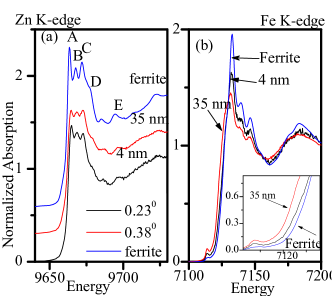
In order to study differences between bulk and surface, we present now a comparison between XANES taken with incidence angle below and above total reflection angle (0.23 degree, attenuation length around 4 nm and 0.38 degree, attenuation length around 35 nm respectively).
In case of ZFO1 (Figure 3), no major changes were observed in the Zn K-edge. The
decrease of peak C could be related to the elimination
of multiple scattering paths due to the high contribution of surface atoms.
The same can be observed for ZFO2 (see Figure 4). In Fe K-edge,
the changes that evidence an increase of Fe in A sites and/or vacancies in
the B site octahedron are less pronounced at the surface. In the case of ZFO5 (Figure 4)
the tendency is similar but the difference with normal spinel is smaller.
Regarding XANES results, we can conclude that in the films grown at low pressure Zn2+ ions are in A sites (coordination four), and Fe3+ are octahedrically (B site) and tetrahedrically coordinated (A site). Also, there is fraction Fe2+ probably in B sites such as in Fe3O4 structure. The increase of Fe3+ in A sites and Fe2+ is favored by the deposition at low oxygen pressure and then due probably to the oxygen vacancy formation. A probe of that is the decrease of features in the XANES Fe K-edge taken with incidence angles lower than total reflection one, i.e. corresponding to the surface region where available environment oxygen neutralize oxygen vacancies. Then, in the case of low pressure growth films there is an increase of Fe3+ tetrahedral sites without inversion.
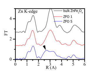
This significant result is supported by EXAFS. Figure 6 shows the Fourier transform (FT) of oscillations extracted from Zn k-edge using Athena program of ZFO1 and ZFO5 films compared with FT of bulk ZnFe2O4 one.
FT of sample ZFO1 is qualitatively equal to bulk ZnFe2O4, reasserting that Zn atoms are in the tetrahedral sites. In the case of ZFO5, the presence of an additional peak around 2.6 A is an indicative that there are Zn atoms at octahedral sites Stewart et al. (2007.).
The situation is different to the scenario found in references Stewart et al. (2007.) and Nakashima et al. (2007): when Zn occupancy of 8-sites increases, Fe decreases, indicating some degree of inversion and magnetic response enhanced with such inversion. In our case the increase of Fe3+ in A sites is not due to Zn and Fe ions inversion, but the mechanism that explains the increase in the magnetization is probably the same as the one reported for inverse spinel. The increase of Fe3+ tetrahedrically coordinated (A site) increases FeA-FeB pairs antiferromagnetically coupled by superexchange, forcing FeB-FeB pairs to align ferromagnetically and also because Fe individual magnetic moments probably no longer cancel. The experimental results report in this work suggest that the increase of Fe3+ in A sites is not due to Zn and Fe ions inversion but of iron situated on normally non-occupied A sites in the spinel structure. In this structure only one eighth of the A sites in a unit cell are actually occupied and so some iron ions at these normally unoccupied sites will act as impurities and will not influence the crystallographic properties.
One more aspect in which oxygen pressure could influence is that as the magnetic interaction in the spinel is an indirect interaction, missing oxygen gives rise to a variation of exchange fields for the ions and a spin/glass like state is formed. Also, the reduction of Fe3+ ions into Fe2+ ions located in octahedral sites would strength ferromagnetic coupling between B-B sites such is the case in magnetite. In the case of Fe2+ located on the A sites there is an increase of magnetic moment due to the antiferromagnetic coupling with Fe3+ but the change will not be as big as for anti-sites.
In summary, we have shown that ferrites grown under low O2 pressure conditions have a large magnetic moment. We found that the inversion mechanism is not responsible for the enhancements of the magnetic interaction – as was found in similar systems – but the presence of defects as oxygen vacancies or iron situated on normally non-occupied A sites in the spinel structure. We have also shown that controlling the oxygen pressure during the deposition, it is possible to obtain conductive or insulating Zn ferrites. These findings allow to control the magnetic and electric transport properties of spinel ferrites controlling the oxygen concentration.
Acknowledgements.
We thank Dr. Silvana Stewart for fruitful discussions and Dr. Azucena Mudarra Navarro for collaboration in XAFS experiments.This work was partially supported by Laboratorio Nacional de Luz Sincrotron, Campinas-Brasil; by CIUNT under Grants 26/E439 by ANPCyT-PICTR 20770 and 35682, by the German-Argentine PROALAR Grant Nr. D/08/11707 and the Collaborative Research Center SFB 762 ”Functionality of Oxide Interfaces”
References
- Yanase and Siratori (1984) A. Yanase and K. Siratori, J. Phys. Soc. Jpn. 53, 312 (1984).
- L ders et al. (2006) U. L ders, A. Barthlmy, M. Bibes, K. Bouzehouane, S. Fusil, E. Jacquet, J. P. Contour, J.-F. Bobo, J. Fontcuberta, and A. Fert, Adv. Mater. 18, 1733 (2006).
- Ayyappan et al. (2010) S. Ayyappan, S. Philip Raja, C. Venkateswaran, J. Philip, and B. Raj, App. Phys. Lett. 96, 143106 (2010).
- Yao et al. (2007) C. Yao, Q. Zeng, G. F. Goya, T. Torres, J. Liu, H. Wu, M. Ge, Y. Zeng, Y. Wang, and J. Z. Jiang, J. Phys. Chem. C 111 (2007).
- Stewart et al. (2007.) S. J. Stewart, S. J. A. Figueroa, J. M. Ramallo Lpez, S. G. Marchetti, J. F. Bengoa, R. J. Prado, and F. G. Requejo, Phys. Rev. B. 75, 073408 (2007.).
- Shim et al. (2006) J. H. Shim, S. Lee, J. H. Park, S. J. Han, Y. H. Jeong, and Y. W. Cho, Phys. Rev. B 73, 064404 (2006).
- Blanco-Gutirrez et al. (2011) V. Blanco-Gutirrez, F. Jimnez-Villacorta, P. Bonvilles, and R. Torralvo-Fernndez, M. J. andSaez-Puche, J. Phys. Chem. C 115 (2011).
- Kamiyama et al. (1992) T. Kamiyama, K. Haneda, T. Sato, S. Ikeda, and H. Asano, Solid State Com. 81, 563 (1992).
- Nakashima et al. (2007) S. Nakashima, K. Fujita, K. Tanaka, K. Hirao, T. Yamamoto, and I. Tanaka, Phys. Rev. B 75, 174443 (2007).
- Chen et al. (2008) Y. F. Chen, D. Spoddig, and M. Ziese, J. Phys. D: Applied Phys. 41, 205004 (2008).
- Lotgering (1966) F. K. Lotgering, J. Phys. Chem. Solids 27, 139 (1966).
- Ligenza (1976) S. Ligenza, Phys. Stat. Sol. 75, 315 (1976).
- Kundu et al. (2003) A. Kundu, C. Upadhyay, and H. Verma, Phys. Lett. A 311, 410 (2003).
- Shenoya et al. (2004) S. D. Shenoya, P. A. Joyb, and M. R. Anantharaman, J. of Mag. and Mag. Mat. 269, 217 (2004).
- Takaobushi et al. (2006) J. Takaobushi, H. Tanaka, T. Kawai, S. Ueda, J.-J. Kim, M. Kobata, E. Ikenaga, M. Yabashi, K. Kobayashi, Y. Nishino, et al., Appl. Phys. Lett. 89, 242507 (2006).
- Kamazawa et al. (2003) K. Kamazawa, Y. Tsunoda, H. Kadowaki, and K. K., Phys. Rev. B. 68, 024412 (2003).
- Fernandes et al. (2009) V. Fernandes, R. J. O. Mossanek, P. Schio, J. J. Klein, A. J. A. de Oliveira, W. A. Ortiz, N. Mattoso, J. Varalda, W. H. Schreiner, M. Abbate, et al., Phys. Rev. B. 80, 035202 (2009).
- Khalid et al. (2009) M. Khalid, M. Ziese, A. Setzer, P. Esquinazi, M. Lorenz, H. Hochmuth, M. Grundmann, D. Spemann, T. Butz, G. Brauer, et al., Phys. Rev. B. 80, 035331 (2009).
- Ramos et al. (2010) M. A. Ramos, J. Barzola-Quiquia, P. Esquinazi, A. Mu oz-Martin, A. Climent-Font, and M. Garca-Hernndez, Phys. Rev. B. 81, 214404 (2010).
- Ohldag et al. (2010) H. Ohldag, P. Esquinazi, E. Arenholz, D. Spemann, M. Rothermel, A. Setzer, and T. Butz, New Journal of Physics 12, 123012 (2010), and refs. therein.
- Sultan and Singh (2009) M. Sultan and R. Singh, J. of Appl. Phys. 105, 07A512 (2009).
- Darko Makovec et al. (2010) D. Darko Makovec, A. Alojz Kodre, I. Iztok Arcon, and M. Miha DrofenikDarko, J. Nanopart. Res. pp. DOI: 10.1007/s11051–010–9929–y (2010).
- Figueroa and Stewart (2009) S. J. A. Figueroa and S. J. Stewart, J. Synchrotron Rad. 16, 63 (2009).
- Akhatar et al. (2009) M. Akhatar, M. Nadeem, S. Javaid, and M. Atif, J. of Phys. Cond. Matt. 21 (2009).
- Als-Nielsen et al. (1994) J. Als-Nielsen, D. Jacquemain, K. Kjaer, F. Leveiller, M. Lahav, and L. Leiserowitz, Physics Report 246 (1994).
- Stoev and Sakurai (1999) K. Stoev and K. Sakurai, Spect. Acta Part B 54 (1999).
- Souza-Neto et al. (2009) N. Souza-Neto, A. Y. Ramos, H. Tolentino, and Y. Joly, J. of Phys.: Conf. Series. 190 (2009).
- Henke et al. (1993) B. Henke, E. Gullikson, and J. Davis, X-ray interactions: photoabsorption, scattering, transmission, and reflection at E=50-30000 eV, Z=1-92, vol. 54 of 2. (Atomic Data and Nuclear Data Tables, 1993), 1st ed., 181-342.