Neutron Imaging by Boric Acid
Abstract
In this paper a new type of passive neutron detector based on the already existing one, CR39, is described. Its operation was verified by three different neutron sources: an Americium-Beryllium (Am241-Be) source; a TRIGA type nuclear reactor; and a fast neutron reactor called TAPIRO. The obtained results, reported here, positively confirm its operation and the accountability of the new developed detecting technique.
1 Introduction
The methods used to detect neutrons are commonly based on the emission of charged particles produced by the interaction of neutrons with He-3 or BF3 contained in the counters or detectors. Another type of commonly used detectors is the BD, the Defender and the DefenderXL which are Bubble Detector-Dosimeters. They are made of an elastic polymer gel in which minute droplets of superheated liquid are uniformly scattered. When these droplets are struck by neutrons, they evaporate and form small bubbles of gas that remain trapped in the elastic polymer. The number of bubbles is in direct proportionality with the number and the energy of neutrons, in particular from the counted number of bubbles in these detectors and from their certified efficiency (Bubbles/mSv) one can obtain the dose equivalent of the neutrons that struck the detector. A further technique is based on CR39. The CR39 (PADC) are plastic detectors made of the polymer C12H18O7, whose density is 1.3 g cm-3. They can detect both heavy charged particles and neutrons when the latter are converted into alpha particles by the nuclear reaction B-10 ( n, ) Li-7 that takes place within a very thin homogeneous layer of B-10 that coats the surface of the CR39 [1]. These detectors have a very wide range of energy extending from tens of KeV (40 KeV) up to some MeV (4MeV). A further technique to measure neutrons is by activation of targets made of thin plates of ultra-pure Au, In, Co, Cu, Mn, Ni, Al, Ti followed by the measurement of the gamma and beta activity of these plates. However, this technique is possible only with quite high fluence of neutrons. The detecting process described in this paper is based on a technique used by the authors in previous experiments in which polycarbonate plates (CR39) were surrounded by a layer of suitable thickness of granular boric acid H3BO3 [2]. In the present case, the polycarbonate plates were substituted for a normal photographic film made of an acetate substrate with a layer of silver halide spread on it. This film was surrounded by a layer of H3BO3 of suitable thickness as it was for the plates. In both cases, the grains of boric acid have the same dimension. Due to a patent pending on this technique we are not allowed to provide further details. This method is based on the same technique described above where neutrons are converted by the boric acid into lithium 7 and alpha particles by the reaction
| (1) |
and the latter interact with a polycarbonate plate on which they produce peculiar tracks.
2 Description and preparation of the detectors
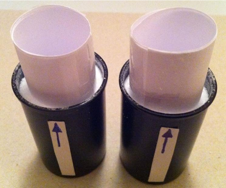
According to the reaction mentioned above, we developed a specific type of detector in which a usual black and white photographic film of 400 ISO made of a layer of silver halide was surrounded by a layer of boric acid. The choice to use a 400 ISO film was made in order to have a compromise between the brightness and the resolution of the image. The detector was made with few frames of photographic film, 24x36 mm, wound in order to form a cylinder by joining the two edges. The second step was to insert this cylindrical film inside a dark cylinder of Polyethylene and then fill this cylinder of boric acid as shown in Fig.1. All of the different tasks to set up the detector were made inside a dark room in complete obscurity, that is without turning on the inactinic light.
3 Experimental Measurements
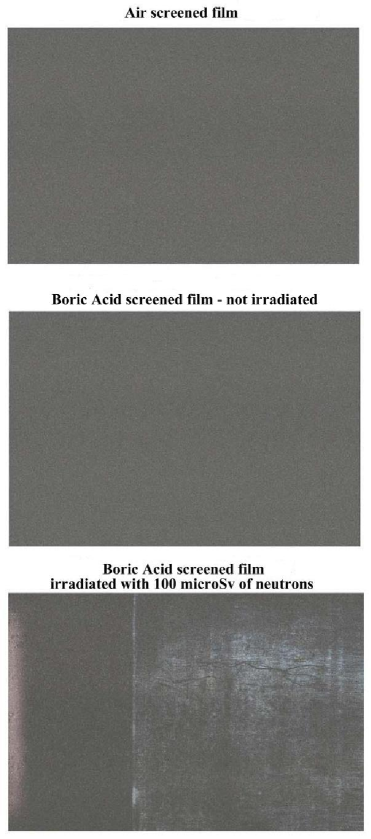
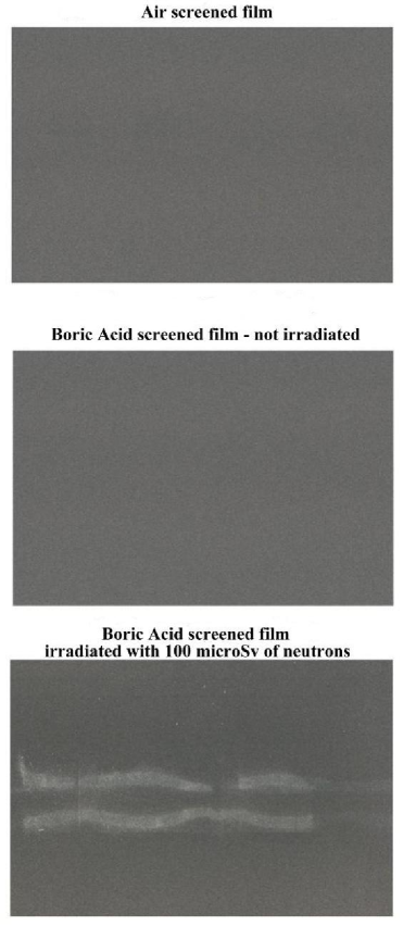
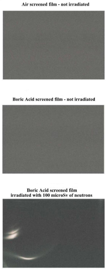
We carried out measurements with three films of the same type described above for each test. In each measurement the three parts of the film were subjected to different conditions: the first part was kept in air and was not irradiated; the second part was kept in boric acid and it was not irradiated; the third part, was immersed in boric acid and was put in front of the neutron flux. These experiments were carried out at the ENEA Casaccia Research Centre, with three types of neutron sources: Am241-Be source; horizontal channel in the thermal column of the nuclear reactor TRIGA; radial channel 2 of the nuclear reactor TAPIRO.
| — | Source Am-241-Be | TRIGA RC1 | TAPIRO |
| neutron | 389 n cm-2 sec-1 | — | — |
| Therm horiz channel | — | 194 n cm-2 sec-1 | — |
| Radial channel 2 | — | — | 278 n cm-2 sec-1 |
| RCd | — | 2.2 | — |
| fixed dose rate (Sv/h) | 100 | 100 | 100 |
| available channels | — | 12 | 7 |
| maximum power | — | 1 MW thermic | 5 kW thermic |
| cooling system | — | Water convection | Helium |
| nuclear fuel | — | Enriched U 20 | Enriched U 93.5 |
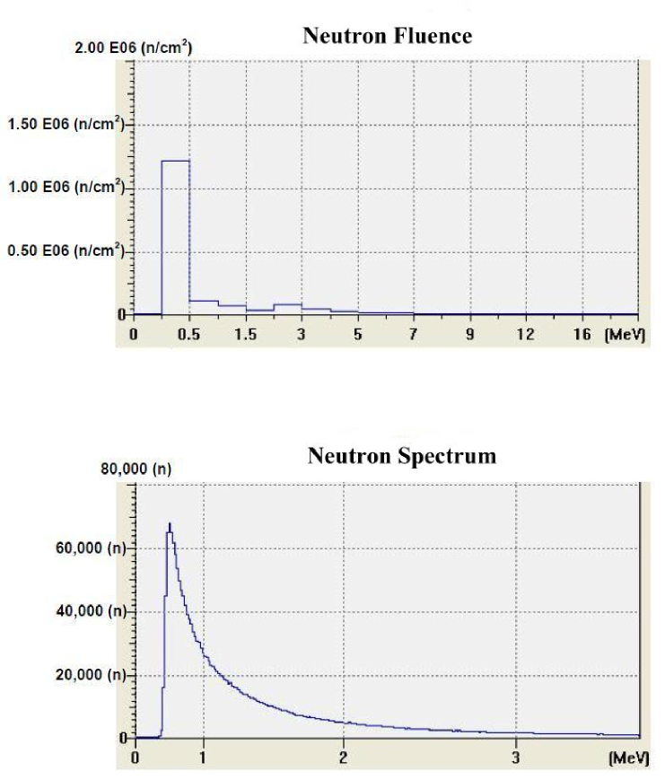
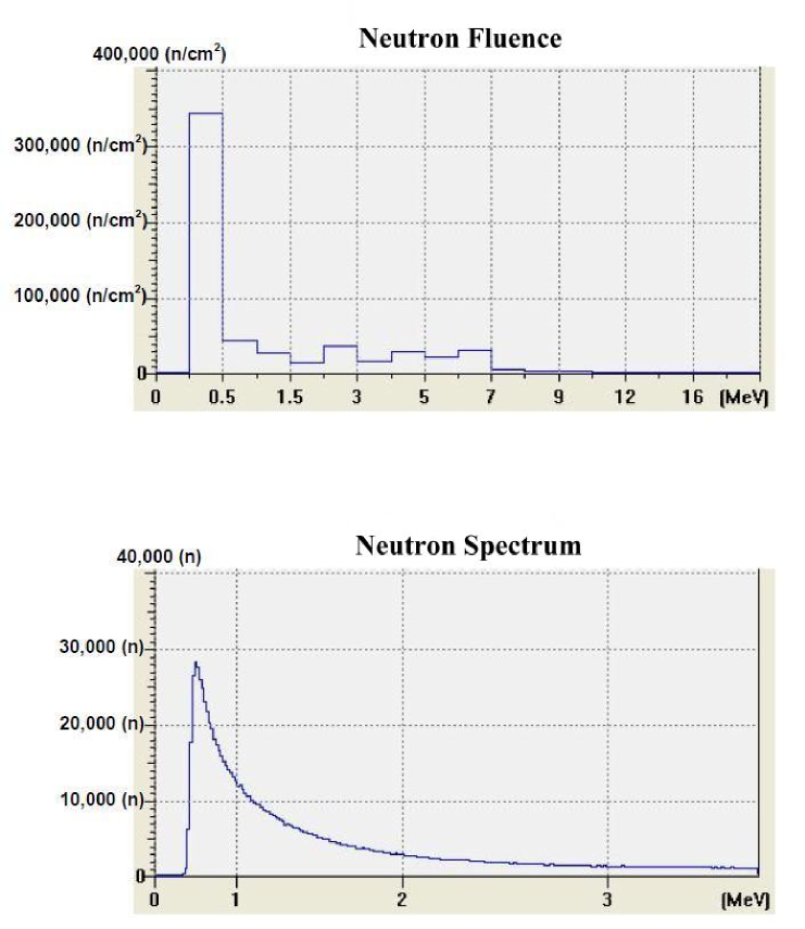
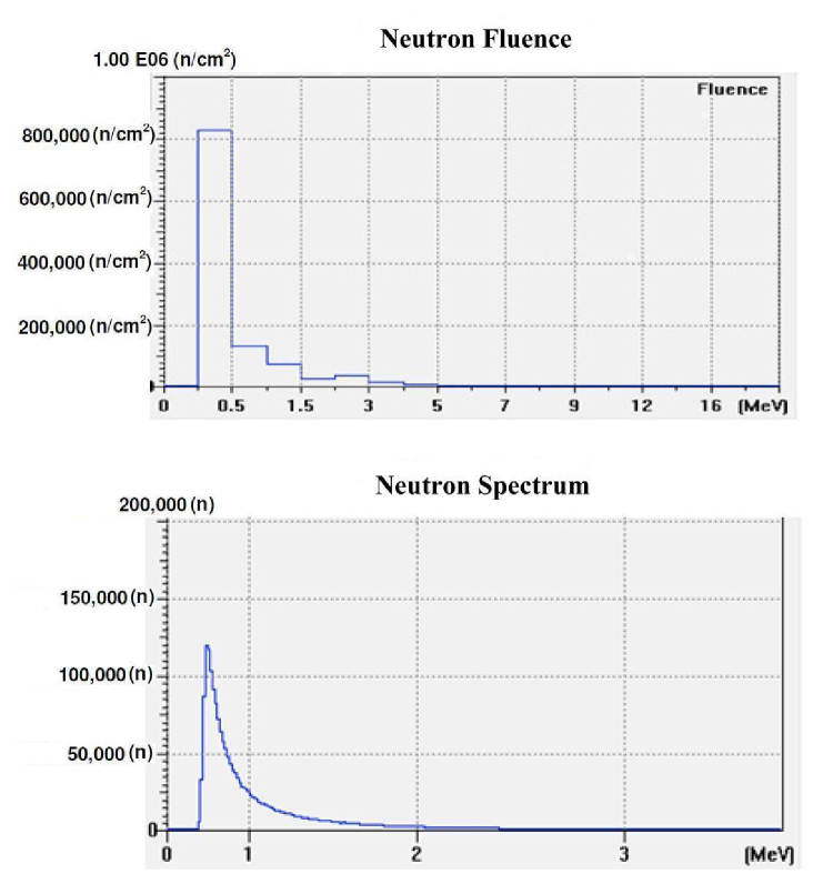
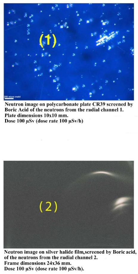
The irradiation time was 1 hour (3600 s) and the dose rate of neutrons was 100 Sv/h for all of the irradiated film. The dose rate had been previously ascertained by an He-3 dosimeter which was placed in the same position where the boric acid detectors will have been placed. In Fig.2 we report the characterisation of the Am-241 - Be source as to the fluence of its neutrons and its neutron spectrums. The characterisation was carried out by the neutron spectrometer MicroSpec-2 [3] Neutron Probe [4] (Bubble Technology Industry) which comprises two detectors ( He-3 and liquid scintillator NE213 ) for two neutron energy intervals. In Figs.3 and 4 we report the fluence and the spectrum, which were measured by the same spectrometer, of the neutrons from the reactors TRIGA and TAPIRO. In all the three measurements, the films with H3BO3, after development, presented visible and clear traces which, conversely, were present neither on the control film screened by H3BO3, nor on the control film in air that were not irradiated. By comparing this experience with a previous one in which we irradiated, in similar conditions, CR39 plates with neutrons in front of the radial channel 2 of the reactor TAPIRO, we find out a very peculiar similarity between the traces obtained on the CR39 screened by boric acid and those obtained on the silver halide of the film screened by boric acid. This detecting technique produces different images for different neutron sources, but for the same neutron source, the images are the same independently of the type of detector used (CR39 or film with silver halide). At the end of the irradiations, the boric acid that had played the role of converting material (it converted neutron into alpha particles) for the irradiated detectors or of a simple screen for the non irradiated ones, was analysed by mass spectrometry. This analysis was conducted in order to look for possible traces of the neutron-into-alpha converting reactions (see 1). In these reactions, the isotope of Boron with higher capture cross section for thermal and slightly epithermal neutrons is B-10. When it captures a neutron, it splits yielding an alpha particle and a nucleus of Lithium-7. Thus, by mass spectrometry we searched both for a possible change in the natural occurring ratio of Boron-11 and Boron-10, expecting a slight decrease of the lighter isotope and a corresponding increase of Lithium-7.
4 Results
Each of the Figs.5, 6 and
7 comprises three images: the first is the
non irradiated film in air; the second is the non irradiated film
immersed in boric acid; the third is the irradiated film screened by
boric acid. In Fig.8, there is the visual comparison
between two images: the tracks on the CR39 and those on the acetate
film with silver halide. These images were obtained by the same
source (reactor TAPIRO) and with the same thickness of boric acid
surrounding the CR39 and the acetate film. Despite the different
substrates (polycarbonate and silver halide) the morphology of the
tracks is the same. With regards to the analyses by mass
spectrometer, we found out a fairly significant variation of the
ratio between B-11 and B-10 in the detectors irradiated by neutrons.
In particular, we found out the two following ratios between
Boron-11 and Boron-10 for the non-irradiated Boron and for the
irradiated one, respectively.
= 3.9906 410-4 non irradiated
= 3.9981 410-4 irradiated
()irradiated ()non-irradiated
Despite this piece of evidence, nothing is possible to say about the presence of Lithium since, even with a significant variation of B-10, the corresponding variation of Lithium-7 was below the minimum value that could be discerned by the mass spectrometer.
5 Conclusion
We developed a new and cheap technique which is capable to detect neutron emission and we verified its accountability by testing it with different sources. These tests produced different images for different sources possessing the same dose rate (Figs.5, 6 and 7). Besides, the same neutron source with equal screening thickness of boric acid surrounding the revealing substrate, produces similar images on different substrates (Fig.8) independently of the revealing substrate (polycarbonate and silver halide). A further proof of the accountability of this technique is available in Fig.6 where the third picture shows the shape of the neutron collimating horizontal slit at the end of the thermal column channel.
6 Aknowledgements
We want to thank for their kind, precious and active collaboration the following people: Giovanni Silvestri owner of the Hobby Photo Laboratories, based in Sulmona (Italy) for the preparation of the film and its development; Massimo Sepielli managing director of the UTFISST ENEA; the team of the nuclear reactor TRIGA of the ENEA Casaccia Research Centre, Daniele Baiano (BAS IONIRP ENEA), Monica Lammardo (UTFISST-REANUC ENEA)reactor operator, Emilio Santoro (UTFISST-REANUC ENEA) director of the reactor TRIGA ; the team of the nuclear reactor TAPIRO of the ENEA Casaccia Research Centre Barbara Bianchi (UTFISST-REANUC ENEA), Orlando Fiorani (UTFISST-REANUC ENEA) and Alfonso Santagata (UTFISST-REANUC ENEA).
References
- [1] R.V. Griffith, D.E. Hankins, L. Tommasino, M.A.M. Gomaa, (United States Patent 4,381,454, April 26th, 1983). ).
- [2] F. Cardone, G. Cherubini, A. Petrucci Phys Letts A 373 (2009) 862.
- [3] www.bubbletech.ca/radiationspectrometersfiles/mobile.html
- [4] www.bubbletech.ca/radiationspectrometersfiles/neutronprobe.html