A Touch of Sleep:
Biophysical Model of Contact-mediated Dormancy of Archaea by Viruses
Abstract
The canonical view of the interactions between viruses and their microbial hosts presumes that changes in host and virus fate require the initiation of infection of a host by a virus. That is, first virus particles diffuse randomly outside of host cells, then the virus genome enters the target host cell, and only then do intracellular dynamics and regulation of virus and host cell fate unfold. Intracellular dynamics may lead to the death of the host cell and release of viruses, to the elimination of the virus genome through cellular defense mechanisms, or the integration of the virus genome with the host as a chromosomal or extra-chromosomal element. Here we revisit this canonical view, inspired by recent experimental findings of Bautista and colleagues (mBio, 2015) in which the majority of target host cells can be induced into a dormant state when exposed to either active or de-activated viruses, even when viruses are present at low relative titer. We propose that both the qualitative phenomena and the quantitative time-scales of dormancy induction can be reconciled given the hypothesis that cellular physiology can be altered by contact on the surface of host cells rather than strictly by infection. We develop a biophysical model of contact-mediated dynamics involving virus particles and target cells. We show how in this model virus particles can catalyze – extracellularly – cellular transformations amongst many cells, even if they ultimately infect only one (or none). We discuss implications of the present biophysical model relevant to the study of virus-microbe interactions more generally.
I Introduction
Dormancy is ubiquitous in microbial systems. One prominent example is that of bacterial persistence, in which cells undergo a rapid physiological change characterized by slowed or even halted growth as well as decreased susceptibility to antibiotics (balaban_2004, ; lewis2007persister, ; lewis_annrev2010, ). Another example is that of microbial “seed banks” (lennon2011microbial, ), in which individual microbes undergo a long period of stasis in which growth is halted. These cells can then restart growth given changes in environmental conditions. A third example is the starvation-dependent division of division of the bacteria Sinorhizobium meliloti into distinct daughter phenotypes, one that is better suited to long-term starvation (ratcliff_2010, ). Long-standing theory (cohen_1966, ) and recent experiments (beaumont_2009, ) support the consensus that varying phenotypes - whether by stochastic bet hedging or via phenotypic plasticity - is an evolutionarily favorable strategy in the face of uncertainty in environmental selection pressures (kussell_sci2005, ). One of the selection pressure that microbial cells face is the possibility of infection and lysis by viruses. Indeed, it has been suggested that E. coli may enter the persistence state as a route to diminish, temporarily, the ability of viruses to eliminate a local population (pearl_2008, ). Nonetheless, the transition between active growth and persistence was not proposed to be a function of virus-host interactions.
Here, we propose a biophysical model of virus-induced dormancy of microbial host cells. Our model is inspired by recent empirical findings by Bautista and colleagues (bautista_inpress, ) of the interactions between the archaeon Sulfolobus islandicus and the dsDNA fusellovirus Sulfulobus spindle shaped virus (SSV9). S. islandicus is a globally distributed archaeon, commonly found in hot spring ecosystems. S. islandicus is also a model system for studying the eco-evolutionary basis for diversity in archaea (zhang2013sulfolobus, ; ISI:000282210700006, ; reno_2009, ; ISI:000262515800015, ; prangishvili_2006, ).
A recent study of the interactions between the host strain S.islandicus RJW002 and the virus SSV9 were recently shown to lead to a novel population-level outcome: “challenge of RJW002 with SSV9 induced a population-wide stasis or dormancy response, where the majority of cells are viable but not actively growing” (bautista_inpress, ). Dormant cells appear “empty” without coherent intracellular structure in contrast to normal cells. Dormant cells can then reorganize and revert to actively growing cells. In the experiment, viruses were introduced at low concentrations relative to that of hosts. Yet, after 24 hrs, nearly 100% of cells were classified as dormant (bautista_inpress, ). In other words: there was a large-scale amplification in the number of dormant cells at the end of the 24 hr period vs. the number of viruses at the start of the experiment. Further, in a follow-up experiment, nearly 100% of cells initiated dormancy even when the host was exposed to de-activated viruses at low relative concentration.
Bautista and colleagues (bautista_inpress, ) highlight the potential role of dormancy as a strategy to survive viral infection and lysis. These experiments also raise the possibility that contact between viruses particles and host surface may be sufficient to initiate a large-scale physiological response, both at the cellular and population-scales. The model we propose here focuses on the early dynamics of virus-host interactions. We assume that viruses can “contact” host cells reversibly. If most contacts between virus particles and host cells do not lead to infection, then there can be a broad dynamic regime in which nearly all of the target host cells enter dormancy even when there are far fewer viruses than hosts. We reach this conclusion by first proposing a biophysical model of contact-mediated dormancy. Next, we solve the model and identify critical transitions between distinct qualitative regimes, including a broad regime of dormancy-enhancement. Finally, we apply the model to the experimental conditions underlying the recent study of RJW002 and SSV9 and show that a contact-model of dormancy is compatible with observations. We discuss the implications of the current work for updating the dominant virus-host infection paradigm to include renewed focus on the role of virus particles in stimulating changes amongst microbes and microbial populations.
II Results
II.1 Biophysical model of contact-mediated dormancy of hosts by viruses
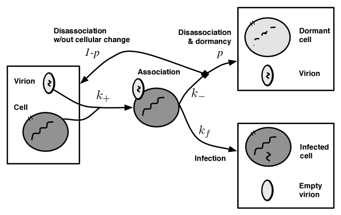
We propose a nonlinear dynamics model of virus-host interactions (see Figure 1). This model describes the early dynamics of interactions involving viruses and hosts and the initiation of either dormancy or an active infection. Consider an environment containing susceptible cells, , and free virus particles, . Free viruses can contact cells forming a complex, , given a diffusion-limited contact rate of cells/(mlhrs). The use of the term “complex” suggests an analogy to models of enzyme kinetics. The complex is reversible. The disassociation rate is cells/(mlhrs) and the infection rate is hrs-1. If disassociation takes place, then the virus is released back into the environment. We assume that disassociation may also induce a cellular transformation leading to dormancy with probability . If infection takes place, then the virus genome enters the host cell, leading to an actively infected cell. The dynamics of this model can be written as:
| (1) |
The system can be reduced in complexity. First there is a constraint that , because the dynamics in the model track the transformation of an initial population of susceptible cells into four different states: susceptible, complex, dormant, and infected. There is another constraint that , because the dynamics track the transformation of an initial population of virus genomes into three different states: in free viruses, temporarily bound with cells, and injected into hosts. Finally, when contact occurs rapidly, then we can use a standard assumption in enzyme kinetics theory and presume that the concentration of rapidly equilibriates (for example, see the Appendix of alon_2007 ). This is a standard approach to analyzing models characterized by fast-slow dynamics. Here, we assume that the change in is relatively fast when compared to other state variables. In the fast limit, then . This approximation is referred to as quasi-steady state approximation (QSSA). Substituting the QSSA equilibrium for the concentration of the complex yields the following reduced system:
| (2) |
We can then identify the following control parameters: the conditional probability of infection given contact, , the effective adsorption rate, , and the ratio of dormancy induction to infection, . Using these control parameters, we can rewrite the model as:
| (3) | |||||
| (4) |
This model can be interpreted as follows. The density of susceptible hosts decreases at a rate proportional to the densities of virus and susceptible host populations (Eq. (3)). The proportionality constant is , the adsorption rate, multiplied by an enhancement factor of , where is the number of dormant cells induced for each infected cell produced. The enhancement factor arises due to the fact that susceptible hosts can become infected or enter dormancy due to interactions with viruses. The number of free viruses decreases at a rate proportional to the densities of virus and susceptible host populations (Eq. (4)). The proportionality constant in that case is , the adsorption rate, because that is the means by which free viruses are removed from the medium.
The model does not include birth and death of hosts, the lysis of hosts by viruses, nor the decay of virus particles. As such, the model describes early dynamics of virus-host interactions. This focus is in contrast to models of virus and host dynamics mediated by density-dependent infection and lysis (levin_1977, ; weitz_2005, ; smith_2012, ; childs_2012, ).
II.2 Qualitative regimes of dormancy induction
Eqs. (3)–(4) can be solved analytically (see Appendix A), yielding:
| (5) | |||||
| (6) |
where and . These solutions hold so long as . When then
| (7) | |||||
| (8) |
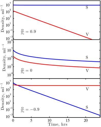
The system dynamics have qualitatively different behaviors for and for (see Table 1). Therefore acts as a critical parameter, both in a biological and dynamical systems sense.
Recall that is the maximum number of hosts that can be infected or enter dormancy as a result of interactions with viruses. Therefore, when then there are enough hosts for all viruses to infect cells ( in total) and to catalyze hosts per infected cell to enter dormancy ( in total). This is the case when is positive. In this limit, all viruses infect a cell, while some hosts remain uninfected. The condition represents the “virus-depletion” limit. In contrast, when there are not enough hosts for all viruses to infect cells and to catalyze hosts per infected cell to enter dormancy. This is the case when is negative. In this limit, all hosts are either infected or enter dormancy, while the viruses remain in the system. The condition represents the “host-depletion” limit. The condition represents the critical point dividing these two dynamical regimes (see Figure 2).
| Variable | |||
|---|---|---|---|
| 0 | 0 | ||
| 0 | 0 | 0 | |
| 0 | 0 |
II.3 Viruses can induce nearly all hosts to enter dormancy, even when the virus-host ratio is far less than one
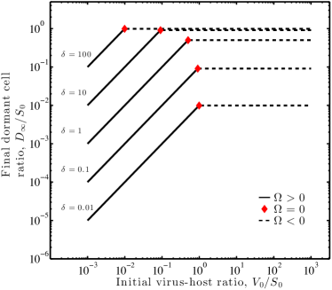
Traditional analysis of virus-host interactions presupposes that entrance, through injection or other means, of virus genomes into a host is required for virus-mediated modification of host cell physiology. Here, as in traditional models, represents the upper limit to the number of hosts infected by viruses (see Table 1). This limit holds when restricting attention to short-term dynamics before replication and lysis which releases more viruses that can initiate subsequent infections. However, in the present model, hosts can also undergo contact-mediated dormancy. When the ratio of viruses to hosts is small, i.e., the MOI is , we find an unexpected outcome: nearly all of the hosts can enter dormancy even when there are far fewer viruses than hosts. In the host-depletion regime, , then . If then . This condition holds so long as the relative rates of unbinding and dormancy are high relative to infection and there are enough viruses. The critical virus density depends on and is equal to . For virus densities above this value, then the dormant cell fraction will have reached its maximum because the system moves from being host-depleted to virus-depleted. The comparison of the asymptotic dormant cell fraction and infected cell fraction are shown in Figure 3. As is apparent, more cells become dormant and infected with increasing titer. Yet, the balance of dormancy or infected cell fates shift with increases in . As increases, then many dormant cells are initiated for each infected cell, whereas when decreases, then very few dormant cells are initiated for each infected cell.

II.4 Dynamics of dormancy induction in the archaeon Sulfolobus islandicus
We apply our biophysical model of contact-mediated dormancy to a recent empirical study of interactions between the archaeon S. islandicus and the dsDNA fusellovirus Sulfulobus spindle shaped virus (SSV9). In the experiment, viruses were introduced at relatively low concentrations compared to that of hosts. The ratio of viruses to hosts, was estimated to range between 0.01, given plaque-forming unit counts, and 0.01, given quantitative PCR counts. In this experiment nearly 100% of host cells entered dormancy. Hence, there was a 10-fold to 100-fold increase in the conversion of host cells into a dormant state. SSV9 was then exposed to UV light to de-activate the virus population. The subsequent conversion estimates into dormant cells were statistically unchanged. We interpret this result to mean that contact between viruses and host may be sufficient to induce a population-wide dormancy response even when viruses are present at densities far below that of target host cells. These qualitative results are the basis for our quantitative parameterization and analysis of the model.
The governing parameters of the biophysical model are , the adsorption rate, and , the ratio of dormancy induction to infection. Bautista and colleagues estimated to be 8.4 ml/min based on the decay of plaque-forming units. They estimated the adsorption rate using the formula where is the original titer of viruses, is the titer at time and is the original titer of hosts. Conventional estimates are that , hence we downward adjust the adsorption rate to be ml/hr (note the change in units). The effective adsorption rate is a combination of the process of diffusion-limited contact and successful infection. We estimate the diffusion-limited contact rate based on physical principles standard in the study of virus-host interactions (bergpurcell_1977, ; murray_meps1992, ):
| (9) |
where is the effective radius of the virus, is the effective radius of the host, where the prefactor is appropriate for interactions taking place at room temperature (293 degrees K) and in a medium with the viscosity of water. Assuming m and m then we predict ml/hr. The ratio of the diffusion-limited contact rate expected from first principles, , and the realized adsorption rate measured in the experiment, can be used to estimate . In this limit, then , such that .
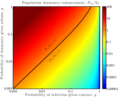
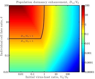
We can explore the predicted fraction of dormant cells as a function of . First, we analyze the ratio of dormant cells to initial viruses given variation in and (Figure 4-left). For there is a broad range of values of such that . This result means that many hosts cells could enter dormancy given exposure to low relative titer of viruses. Yet, the value of , and therefore of , remains a free parameter given the experimental tests conducted in the system. The maximum value of is when , i.e., when the conditional probability of inducing dormancy given a reversible contact approaches 1. In this case, . Lower values of are possible when , i.e., when the conditional probability of inducing dormancy given a reversible contact approaches 0. In this limit, . The experimental finding of a range between to fold enhancement is consistent with our finding of and a model in which (see Figure 4-right).
We provide another evaluation of the model by considering the timescale over which contact-mediated dormancy should take place. The appropriate time-scale is predicted to be . The approximate cell density for experiments in (bautista_inpress, ) was ml-1. We assume that viruses were present at . Using these values we estimate hrs. Hence, we predict a characteristic time-scale for conversion of 64% of hosts, corresponding to a one-log drop in susceptible host density, in a time period of 4 hrs and to conversion of 87% of hosts, corresponding to a two-log drop in susceptible host density, in a time period of 8 hrs. We view this time-scale analysis to be another confirmation of the model, given that even if the hosts initiate dormancy after contact, the dormancy “phenotype” is likely to be delayed given the re-organization of intracellular dynamics. In summary, non-infectious contacts could happen sufficiently frequently so as to rapidly induce dormancy on relevant time-scales of experimental observations.
III Discussion
We have proposed a biophysical model of host-dormancy initiated by contact with viruses. The model explicitly accounts for the possibility that viruses can contact host cells reversibly. Reversible contacts may, with some frequency, lead to induction of dormancy. Such contact-mediated dormancy at the cellular scale can be evident at the population-scale in certain limits. In particular, we predict a critical transition to a regime in which the vast majority of cells become dormant even if the initial ratio of viruses to hosts is quite small. This regime is found to be robust to a broad range of biophysically-relevant parameters.
The inspiration for the model was a recent series of findings that the majority of an archaeal population could enter a stasis-like “dormancy” in less than 24 hrs after exposure to a relatively small number of viruses bautista_inpress . The same effect was observed whether active or inactivated viruses were utilized. This experimental finding suggests the possibility that contact between virus particles and host surfaces induce a transformation in host phenotype. Dormant cells were unlikely to be infected and lysed by viruses. However, such dormancy comes at a cost, as residing in a dormant state for too long can lead to loss of cell viability and cell death (bautista_inpress, ). This transformation may represent a form of phenotypic plasticity on the part of hosts. Further work would be required to evaluate whether dormancy could be initiated independent of virus-contact, which would represent a form of bet hedging.
We fit our model to the experimental host-virus system, leaving only one free parameter: the conditional probability of dormancy initiation upon a reversible contact. We predict that whenever this conditional probability is sufficiently high, then large-scale initiation of dormancy can occur even when very few cells are infected. Based on our fits, we predict rapid initiation of dormancy can take place on a time-scale of 4-8 hrs, sufficiently fast so as to identify a dormancy phenotype amongst the majority of the host population in the 12-24 hrs period as observed. Moreover, the same model fits predicts the potential for a 50-fold enhancement in dormant cells with respect to viruses. Experiments observe higher enhancement ratios ranging from 10- to 100-fold (bautista_inpress, ). The uncertainty is due, in part, to challenges in quantifying virus titer. Yet, there are other challenges, including potential errors in the estimation of the adsorption rate and approximation of the maximum contact rate. There may also be additional mechanisms of relevance, e.g., cell-cell communication, as a means to amplify a small viral contact “signal” or additional spatial structure of the cells and viruses in the environment not accounted for in the present model. Nonetheless, the current biophysical model provides an explanation for the qualitative features of dormancy-enhancement and the time-scale of the effect. We suggests that the present baseline model serve as the basis for future detailed investigations of the phenomena moving forward.
A number of issues remain to link the proposed early-time dynamics with long-term dynamics. First, nonlinear feedbacks are likely to arise in this system due to the infection and release of viruses. These viruses are themselves metastable, and so incorporating the decay of infectious virus particles will also need to be considered. Second, here we assume that viruses cannot infect dormant cells. The interaction between viruses and dormant cells is not fully elucidated. Finally, it is known that intracellular interactions of S. islandicus and its viruses are mediated, in part, by the CRISPR/Cas immune system. The CRISPR/Cas immune system is ubiquitous in bacteria and archaea. CRISPR/Cas enable host cells to target and degrade foreign genetic elements, including viruses (barrangou_2007, ; horvath_2010crispr, ; vanderoost_2014, ). CRISPR-mediated interactions can lead, over time, to the diversification of the host as it obtains new immune elements from the virus and to the diversification of the virus (andersson_2008virus, ; isi_:000282210700006, ; weinberger_2012, ; held2013reassortment, ). Linking early- to long-term dynamics will also need to confront the potential diversification of communities arising due to contact-mediated and infection-mediated dynamics.
In summary, dormancy is a feature of organisms spanning animals to plants to microbes. The evolution of dormancy has long been thought to represent a way to maximize long-term fitness in an uncertain environment (cohen_1966, ). For microbes, part of the uncertainty in their fitness stems from the possibility that they may be infected and lysed by a virus. Here, we find that a biophysical mechanism of context-initiated dormancy can explain observations of rapid and large-scale conversion of a host archaeal population into a dormant state by a relatively small number of viruses. We suggest that detailed investigations of contact of hosts by viruses is likely to yield new biological surprises. In turn, new theoretical approaches are needed to consider the integration of “fast” dynamics at contact-scales with the long-term nonlinear feedbacks arising from the effects of physiological transformations and infection on host and virus populations.
Acknowledgments
The authors thank M. Bautista, R. Whitaker and M. Young for helpful comments and feedback on the manuscript. JSW acknowledges support from a Career Award at the Scientific Interface from the Burroughs Wellcome Fund and NSF Award 1342876.
Appendix A Analytical solution of the contact-mediated dynamics model
The following reduced model introduced in Eqs (3)–(4) represents dynamics of susceptible hosts, , and free viruses, :
| (10) | |||||
| (11) |
This model can be solved analytically by observing that:
| (12) |
such that
| (13) |
where the integration constant, , can be solved using the initial conditions that and :
| (14) |
This two-dimensional system can be reduce to a one-dimensional system by substituting as in Eq. (13), yielding:
| (15) |
When , then Eq. (15) can be solved by separation of variables yielding:
| (16) |
where is the initial population-level multiplicity of infection, i.e., the ratio . The solution for is the basis for a complete description of the dynamics under the Quasi-steady state approximation:
| (17) |
where, in addition:
| (18) |
When , then Eq. (15) can be solved by integration:
| (19) |
recalling that when then .
References
- (1) Balaban NQ, Merrin J, Chait R, Kowalik L, Leibler S (2004) Bacterial persistence as a phenotypic switch. Science 305:1622–1625.
- (2) Lewis K (2007) Persister cells, dormancy and infectious disease. Nature Reviews Microbiology 5:48–56.
- (3) Lewis K (2010) Persister cells. Annual Review of Microbiology 64:357–372 PMID: 20528688.
- (4) Lennon JT, Jones SE (2011) Microbial seed banks: the ecological and evolutionary implications of dormancy. Nature Reviews Microbiology 9:119–130.
- (5) Ratcliff WC, Denison RF (2010) Current Biology pp 1740–1744.
- (6) Cohen D (1966) Optimizing reproduction in a randomly varying environment. Journal of Theoretical Biology 12:119 – 129.
- (7) Beaumont HJE, Gallie J, Kost C, Ferguson GC, Rainey PB (2009) Experimental evolution of bet hedging. Nature 462:90–93.
- (8) Kussell E, Leibler S (2005) Phenotypic diversity, population growth, and information in fluctuating environments. Science 309:2075–2078.
- (9) Pearl S, Gabay C, Kishony R, Oppenheim A, Balaban NQ (2008) Nongenetic individuality in the host-phage interaction. PLoS Biology 6:957–964.
- (10) Bautista MA, Zhang C, Whitaker RJ (2015) Virus-induced dormancy in the archaeon Sulfolobus islandicus. mBio 6:e02565–14.
- (11) Zhang C, Krause DJ, Whitaker RJ (2013) Sulfolobus islandicus: a model system for evolutionary genomics. Biochem. Soc. Trans. 41:458–462.
- (12) Held NL, Herrera A, Cadillo-Quiroz H, Whitaker RJ (2010) CRISPR Associated Diversity within a Population of Sulfolobus islandicus. PLoS One 5:e12988.
- (13) Reno ML, Held NL, Fields CJ, Burke PV, Whitaker RJ (2009) Biogeography of the Sulfolobus islandicus pan-genome. Proc. Natl. Acad. Sci. USA 106:8605–8610.
- (14) Held NL, Whitaker RJ (2009) Viral biogeography revealed by signatures in Sulfolobus islandicus genomes. Environmental Microbiology 11:457–466.
- (15) Prangishvili D, Forterre P, Garrett RA (2006) Viruses of the archaea: a unifying view. Nat Rev Micro 4:837–848.
- (16) Alon U (2007) An Introduction to Systems Biology: Design Principles of Biological Circuits (Chapman and Hall/CRC, Boca Raton, FL).
- (17) Levin BR, Stewart FM, Chao L (1977) Resource-limited growth, competition, and predation: a model and experimental studies with bacteria and bacteriophage. American Naturalist 111:3–24.
- (18) Weitz JS, Hartman H, Levin SA (2005) Coevolutionary arms races between bacteria and bacteriophage. Proceedings of the National Academy of Sciences of the United States of America 102:9535–9540.
- (19) Smith HL, Thieme HR (2012) Persistence of bacteria and phages in a chemostat. J. Math. Biol. 64:951–979.
- (20) Childs LC, Held NL, Young MJ, Whitaker RJ, Weitz JS (2012) Multi-scale model of CRISPR-induced coevolutionary dynamics: diversification at the interface of Lamarck and Darwin. Evolution 66:2015–2029.
- (21) Berg HC, Purcell EM (1977) Physics of chemoreception. Biophysical Journal 20:193–219.
- (22) Murray AG, Jackson GA (1992) Viral dynamics: a model of the effects of size, shape, motion and abundance of single-celled planktonic organisms and other particles. Marine Ecology Progress Series 89:103–116.
- (23) Barrangou R, et al. (2007) CRISPR provides acquired resistance against viruses in prokaryotes. Science 315:1709–12.
- (24) Horvath P, Barrangou R (2010) CRISPR/Cas, the Immune System of Bacteria and Archaea. Science 327:167–170.
- (25) van der Oost J, Westra ER, Jackson RN, Wiedenheft B (2014) Unravelling the structural and mechanistic basis of CRISPR-Cas systems. Nat Rev Micro 12:479–492.
- (26) Andersson AF, Banfield JF (2008) Virus population dynamics and acquired virus resistance in natural microbial communities. Science 320:1047–1050.
- (27) Held NL, Herrera A, Cadillo-Quiroz H, Whitaker RJ (2010) CRISPR Associated Diversity within a Population of Sulfolobus islandicus. PLoS One 5.
- (28) Weinberger AD, et al. (2012) Persisting viral sequences shape microbial CRISPR-based immunity. PLoS Comput. Biol. 8:e1002475.
- (29) Held NL, Herrera A, Whitaker RJ (2013) Reassortment of crispr repeat-spacer loci in sulfolobus islandicus. Environmental microbiology 15:3065–3076.