Solvent Exchange in a Hele-Shaw Cell: Universality of Surface Nanodroplet Nucleation
Abstract
Solvent exchange (also called solvent shifting or Ouzo effect) is a generally used bottom-up process to mass-produce nanoscale droplets. In this process, a good solvent for some oil is displaced by a poor one, leading to oil nanodroplet nucleation and subsequent growth. Here we perform this process on a hydrophobic substrate so that sessile droplets – so-called surface nanodroplets – develop, following the work of Zhang et al. [Zhang, X.; Lu, Z.; Tan, H.; Bao, L.; He, Y.; Sun, C.; Lohse, D. Proc. Natl. Acad. Sci. U. S. A. 2015, 122, 9253-9257]. In contrast to what was done in that paper, we chose a very well controlled Hele-Shaw geometry with negligible gravitational effects, injecting the poor solvent in the center of the Hele-Shaw cell, and characterize the emerging nanodroplets as function of radial distance and flow rates. We find that the mean droplet volume per area strongly depends on the local Peclet number and follows an universal scaling law . Moreover, the probability distribution function of the droplet volume strongly depends on the local as well, regardless of the flow rates and radial distance, giving strong support to the theoretical model of the solvent exchange process developed in Zhang et al’s work.
keywords:
American Chemical Society, LaTeXPhysics of Fluids Group, Department of Applied Physics and J. M. Burgers Centre for Fluid Dynamics, University of Twente, P.O. Box 217, 7500 AE Enschede, The Netherlands \alsoaffiliationBeijing Advanced Innovation Center for Biomedical Engineering, Beihang University, 37 Xueyuan Rd, Haidian District, Beijing, China \alsoaffiliationPhysics of Fluids Group, Department of Applied Physics and J. M. Burgers Centre for Fluid Dynamics, University of Twente, P.O. Box 217, 7500 AE Enschede, The Netherlands \abbreviationsIR,NMR,UV
![[Uncaptioned image]](/html/2002.03030/assets/figs/TOC.png)
1 Introduction
The solvent shifting process – also called solvent exchange – is a simple and generic approach for mass-producing droplets or bubbles at solid–liquid interfaces. The droplet or bubble are only several tens to hundreds of nanometers in height, or a few femtoliters in volume 1, 2, 3, 4, 5, 6. In this process, a good solvent is replaced by a poor solvent, leading to nanodroplets or nanobubble nucleation and subsequent growth on the substrate. The approach has several potential applications, such that it can be used for liquid-liquid microextraction, diagnosis, drug production, extraordinary focusing, micromanufacture and among many others7, 8, 9, 10, 11, 12, 13, 14, 15, 16, 17, 18, 19. When applied to oil dissolved in a good solvent, the solvent exchange process has the capability of mass-producing surface nanodroplets of oil on substrates in one step20, 21, 22, 23, 24, 25.
In ref. 20 the solvent exchange process was performed in a linear channel, with the flow rate and the channel height as control parameters. The nanodroplet generation for seven different flow rates and three different channel heights between mm and mm was analysed. The main result was that the experimentally found mean droplet volume was consistent with the theoretical results derived in the same paper, namely that the area-averaged volume of the droplet , where area-averaged volume is defined by the total volume of droplets over a unit surface area, is the Peclet number, defined by the flow rate, the width of the channel (where is the rectangular channel cross section), and the mass diffusivity of the oil, see figure 2G of that paper.
However, for the larger channel heights analyzed in that paper, major convective effects sets in, due to the density difference between the two solvents, leading to considerable inhomogeneities in droplet sizes. Indeed, the role of gravity in the solvent exchange process in such thick channels could later be confirmed in ref. 26.
The aim of this present paper therefore is to go to a different geometry, namely to a Hele-Shaw cell: a channel formed by two closely spaced parallel glass plates. The employed Hele-Shaw cell possesses a much smaller cell height of , implying an Archimedes number (see ref. 26) of , where is the gravitational acceleration, is the kinematic viscosity, is the density of the fluid for injection (deionized water, in this case), and is the density difference between a solution to be repelled from the Hele-Shaw cell (water-ethanol solution with the ethanol concentration of , in this case) and the fluid for injection. For , gravitational effects can be neglected. The Hele-Shaw geometry has the additional advantage that the flow rate now depends on the radial distance from the point of flow injection, allowing for continuous variation of the local flow velocity, which again leads to the continuous tuning of the Peclet number, which is again the non-dimensionalized flow rate, here given by
| (1) |
As a result, the correlation of nanodroplet formation with Peclet number can be easily investigated. The main question which arises is: Does the relationship also locally hold in the Hele-Shaw geometry and is thus universal?
Figure 1 shows a sketch of the geometry and the employed notations for the nanodroplets. With this setup, we want to test the fluid dynamical theory of solvent exchange (which can straightforwardly be generalized to the present circular geometry) developed in reference 20.
The paper is organized as follows: Section 2 gives the details on the employed method and how the data were collected. Section 3 shows the results, followed by a discussion and the conclusions (section 4).

2 Experimental Section
2.1 Sample preparation
A circular glass cover slip (GOLO, China) with a diameter of 50 mm was used as the substrate for droplet nucleation and bottom window for observation in the Hele-Shaw cell. The substrate was hydrophobilized by PVDF-HFP (poly(vinylidene fluoride-co-hexafluoropropylene), Mw = 400000, Sigma-Aldrich, USA). To do so, the cover slip was first cleaned in a sonication bath of piranha solution (70% H2SO4- 30% H2O2 solution) for 30 min, followed by the sonication bath of acetone and ethanol for 30 min, then in deionized water 3 times for 5 min. After that, the sample was dried by nitrogen gas. The dried cover slip was then immersed in a 3 PVDF-HFP in a petri dish. The petri dish remained in an oven at 150∘C for 12h. Eventually, a hydrophobic glass substrate coated with PVDF-HFP was obtained. The measured static contact angle of water on the obtained PVDF-HFP hydrophobic surface is 110∘.
2.2 Formation of nanodroplets through solvent exchange in the Hele-Shaw geometry
Nanodroplets of trans-anethole (4-Propenylanisole, trans-1-Methoxy-4-(1-propenyl)benzene, Solarbio, USA) were produced on the obtained hydrophobic substrate in a Hele-Shaw cell through solvent exchange. As shown in Figure 1a, the top cover slip and the bottom cover slip window form a disk-shaped channel with a height and a diameter . Unlike the liquid cell applied in ref. 20, the inlet of the Hele-Shaw cell is in the center of the cell. The size and area density of the droplets are influenced by the solution composition for the solvent exchange 24, 27. In our experiments, two solutions, solvent A and solvent B, were prepared for solvent exchange. Solvent A is an aqueous ethanol solution with the ethanol (analytically pure,99.8, Aladdin, China) concentration of . The solution was saturated with trans-anethole, which was labeled by a fluorescence dye perylene (Klamar-reagent, China). Solvent A serves as good solvent for trans-anethole with saturation concentration of . Solvent B is deionized (DI) water, which serves as poor solvent for the trans-anethole. Before the experiments, solvent B was saturated with trans-anethole as well. Such solution composition produced desirable size and density of droplets, suitable for optical images and data analysis. During the solvent exchange process, solution A was displaced by 2 mL solution B. The injection of solution B was performed by a syringe pump at three different flow rates of , , and .
2.3 Characterization of nanodroplets
After the tran-anethole droplets had nucleated on the hydrophobic glass substrate, they were characterized by a reflecting fluorescence microscope (IX71, Olympus, Japan). Two objectives ( and ) were used in the imaging of the nucleated droplets. The images taken with the 20 objective were used for morphological analysis, while the ones taken with the 4 objective were used to determine the radial distance for each selected area in the the optical images taken with the 20 objective. All the optical images () were analyzed using a home designed image segmentation algorithm for the optimized extraction of the droplet footprint diameters. For details of the algorithm, readers are referred to our previous publications 28, 29, 30, 31.
For each of the three flow rates, the nucleated nanodroplets with different radial distance were captured. The number of the captured nanodroplets are 18332, 14403, and 19990 for flow rates of , , and , respectively. For each , the captured droplets were binned in concentric circular bands with a band width .
To extract volumes of the nucleated nanodroplets, a tapping mode atomic force microscopy (TM-AFM) (Resolve, Bruker, USA) was also applied to get high-resolution three dimensional (3D) images of the nucleated droplets, after the growth of the oil droplets was already finished. The nucleated nanodroplets remained in solvent B and were imaged in the liquid mode of TM-AFM. For the duration of the operational time, we did not observed any temporal evolution in the droplet lateral size. The obtained AFM images were analyzed with a home-designed Matlab program to extract the height, width and contact angle of the droplets, as reported in our previous work 32. After that, the volumes of the nanodroplet can be obtained.
Since the number of the captured nanodroplets is huge, it is impractical to measure the volumes of all individual nanodroplets with AFM. Therefore, in this study, only a few nanodroplets with different footprint diameters were imaged with TM-AFM, in order to establish the dependence of the nanodroplet height and contact angle on its footprint diameter. The height of droplets was measured for droplets with different footprint diameter , as shown in figure 3b. From the relationship, we obtained the dependence, as shown in the dotted blue curve, which provides the basis for estimation of the contact angle for droplets with different determined from the optical images. This dependence was further used to obtain the volume for each captured nanodroplet in the optical images from the footprint diameter.
2.4 Fluid dynamics theory of solvent exchange
The scaling law between the final area-averaged volume of the droplet and the Peclet number was established in ref. 20. When gravitational effect in solvent exchange process can be ignored (i.e., for small Archimedes number), the final area-averaged droplet volume scales as:
| (2) |
where and are the saturation concentrations of oil in the poor solvent and the actual oil concentration, respectively, and is the oil density. This fluid dynamics theory of solvent exchange will be further experimentally validated in this paper.
3 Results and Discussion
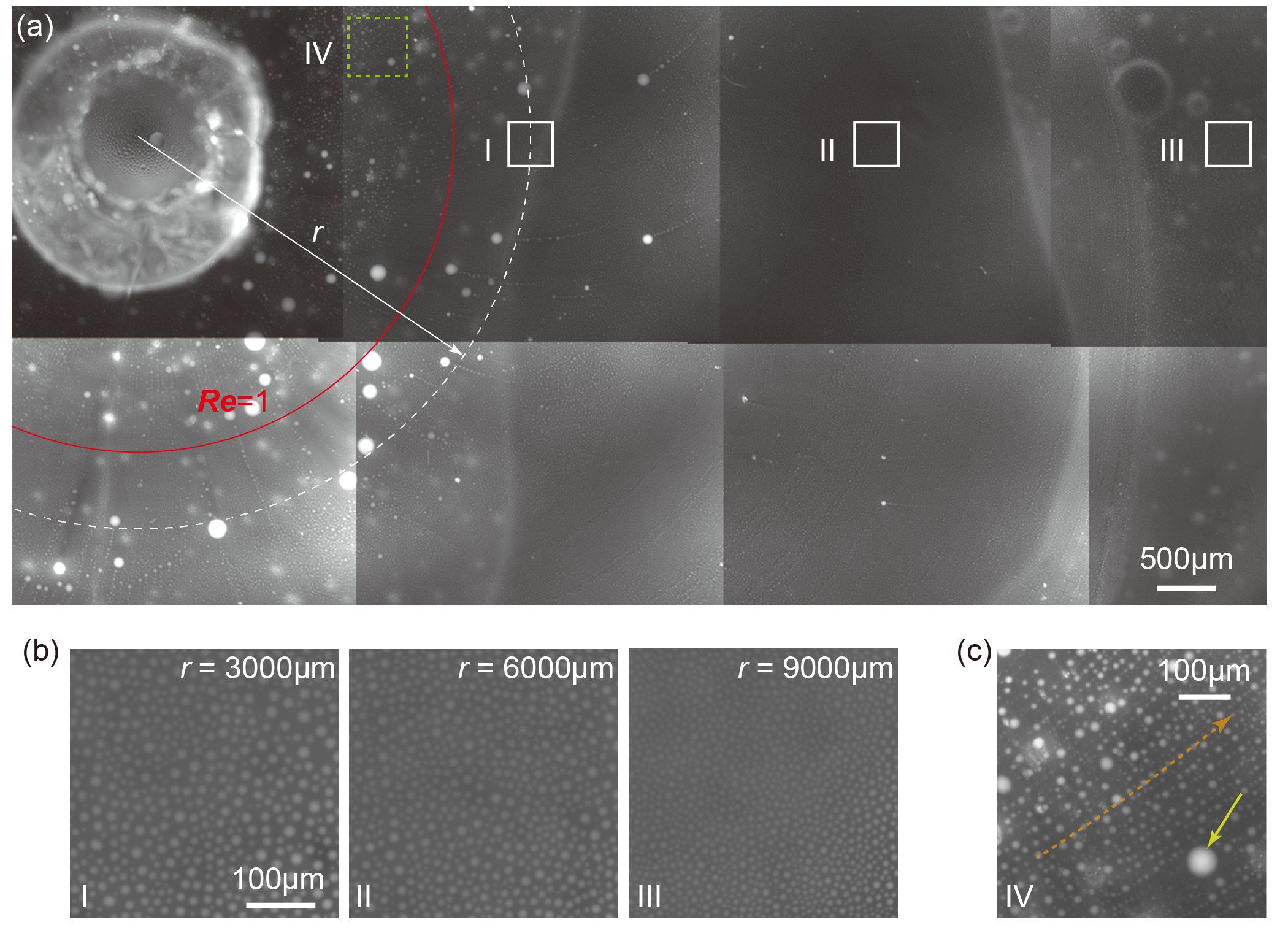
After solvent exchange, nucleated nanodroplets were attached to the bottom cover slip window. Figure 2a shows an fluorescence image obtained by aligning different images taken with the 4 objective at the flow rate of . Three examples of images taken with a higher magnification objective of 20 are shown in figure 2b for the three selected areas in figure 2a. From the images, one can clearly see that the droplet size decreases with increasing distance from the center. Figure 2c shows an enlarged image for an area selected by the dotted green box in figure 2a. One can see that the droplets in the area are lined up along the flow direction (as guided by a dotted orange arrow) and exhibits nonuniform size distributions. This will be discussed later in this section.
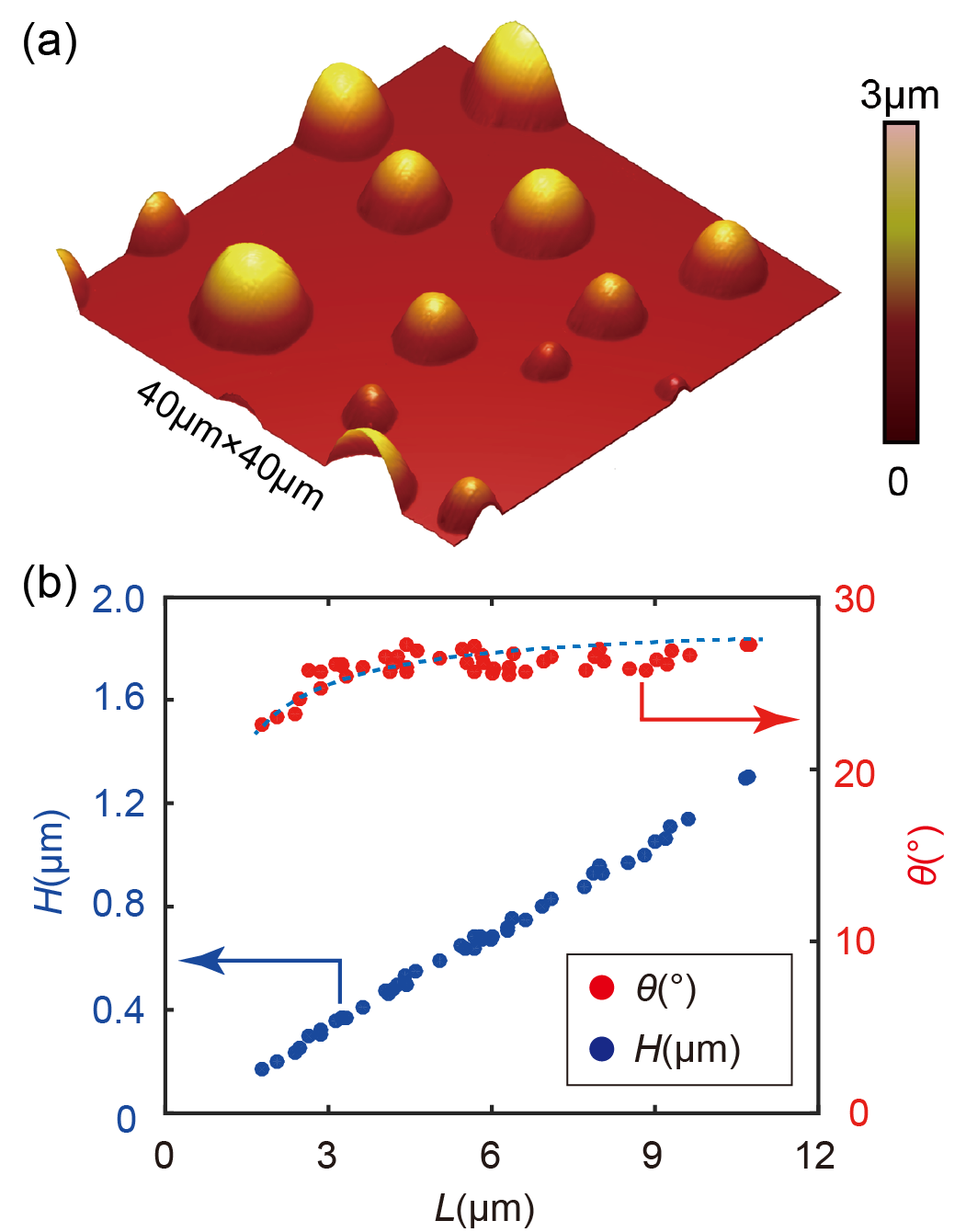
Figure 3a shows a representative AFM image of the trans-anethole droplets scanned in solvent B on the bottom substrate. From the AFM images, the height and the contact angle for the nanodroplets were extracted, as shown in figure 3b. One can see that the droplet height roughly linearly increases from about to as increases from about to . The contact angle basically remains constant at after is larger than . The aspect ratios of the nucleated droplets are consistent with the results reported in ref.33, 20 for the same combination of solvents and solutes.
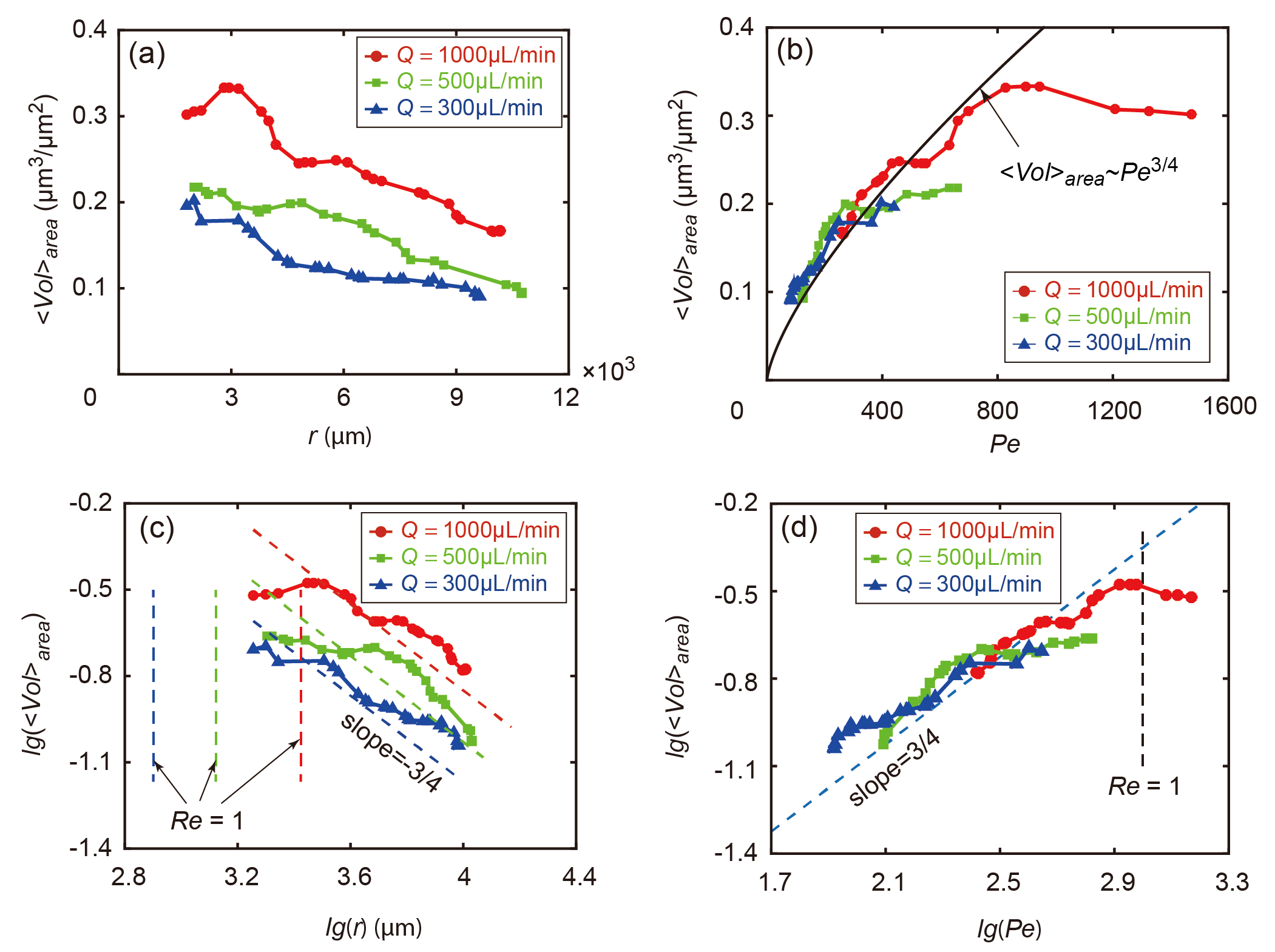
With the developed dependence between , , and , the droplet volumes for individual captured droplets in the optical microscope images were obtained. After that, the mean droplet volume per area area was further calculated, as shown in figure 4a. For all the three flow rates, decreases with increasing . Moreover, for the same , increases with increasing flow rates. Most importantly, regardless of the flow rates, all the three curves are well superposed on each other in the plot of versus Peclet number , as shown in figure 4b, especially for lower value (corresponding to larger distance from the inlet).
The double logarithmic plots of versus the radial distance and vs the Peclet number are shown in figure 4c,d. It is clear that shows a -3/4 and 3/4 power law dependence on and , respectively. Most importantly, three curves in figure 4d collapse on one universal curve. These results are consistent with the theoretical prediction (eq. (2)) and provide strong support to the model.
In figure 4c, d, for smaller or larger , the data points for start to deviate from the -3/4 or 3/4 power law scaling lines. The deviation is likely due to the higher Reynolds number corresponding to smaller . In the Hele-Shaw cell, the Reynolds number at the radial distance is given by
| (3) |
where is the kinematic viscosity of the solution (here for water). In figure 4c, the red, green, and blue vertical dashed lines correspond to for the flow rates of , , and , respectively. In figure 4d, a dashed vertical line was also drown at the position of . One can see that the deviations mainly occur in the area where the Reynolds number . Indeed, at higher Reynolds numbers, the flow becomes dominated by inertia effects and the laminar theory of ref20 would require extensions. Namely, vortical flow structures may occur, leading to a less organized flow pattern. This may lead to advection of nucleated droplets.
For that case , droplets indeed can move outwards along the radial direction of the Hele-Shaw cell. This can be seen from figure 2c. It is an enlarged image for the area highlighted by the dotted green box in the region corresponding to , which is defined by a red circle in figure 2a. In the image, one can see that the droplets are mostly lined up along the radial direction, which is clearly different from that shown in figure 2b. The advected droplets can merge with other surface droplets on their paths, leading to an increased size of droplets, as pointed at by the arrow in figure 2c. Meanwhile, the strong outward motion of droplets unavoidably leads to a reduced average volume close to the center flow inlet. As a result, in the region with , is smaller than what is predicted by the model equation(2) of reference20 and the experimental results deviate from the power law line, as shown in figure 4c,d.
Compared to the result reported in ref. 20 (Figure 2G, therein), the result shown in figure 2d provide a higher consistency among the experiments with different flow rates. This is attributed to the smaller cell height of , which eliminates gravitational effects (i.e., Ar < 1). The results thus give strong support to our previously developed fluid dynamics model of solvent exchange, namely, that the volume of the nucleated droplets strongly depends on the dimensionless flow velocity, perfectly following the relationship .
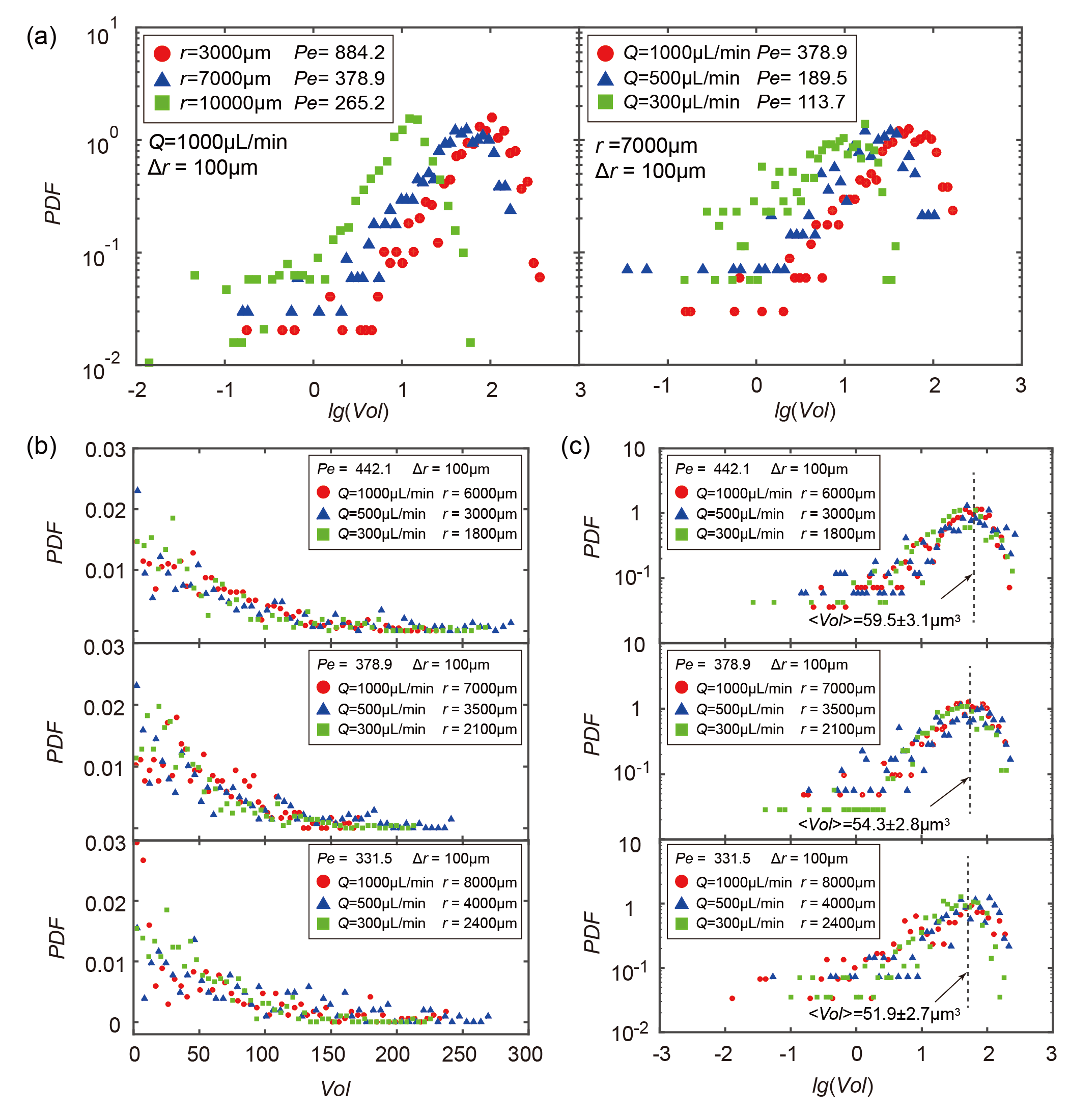
After having shown the universality of the 3/4 power law dependence between and , one also wonders on the universality of the probability distribution function (PDF) of the droplet volumes, i.e., on the dependence of the PDFs on . To answer this question, the distribution of the droplet volume was calculated. Figure 5a shows the PDF of the droplet volume at different for the flow rate (left figure) and at the same radial distance for different flow rates (right figure). In the left figure, the PDF of droplet volumes shifts rightwards with decreasing . Similarly, the PDF shifts rightwards with increasing in the right figure. Both figures of course reflect that the mean droplet volume increases with .
Since is continuously varying in the Hele-Shaw cell, this gives us the freedom to select a particular value for fixed flow rate. This allows us to compare the distribution of the droplet volume with the same value, but different . For the three different flow rates, three different local values of , , and were selected. This corresponds to nine areas in total, three for each of flow rate. The linear and double-logarithmic plots of the PDF of the droplet volumes for the three selected values are shown in figure 5b and c, respectively. Remarkably, it shows that the PDFs of the droplet volume have an universal dependence on the Peclet number, regardless of the radial distances and flow rates .
4 Conclusions
In summary, we experimentally investigated the formation of surface nanodroplets by solvent exchange under a well controlled flow conditions. Compared to the rectangular cross section channels used in one of our previous study 20, a Hele-Shaw cell with a cell height of 100 was employed. In the new setup, gravitational effects can be negligible. Moreover, the Hele Shaw setup easily allows the continuous tuning of the dimensionless flow velocity - namely the Peclet number . By combining a fluorescence optical microscope and an AFM, the height, contact angle, footprint diameter, and volume of surface nanodroplets were extracted for a huge amount of nanodroplets under three different flow rates. The results reveal the underlying mechanism governing the droplet nucleation through solvent exchange. They show that not only the mean droplet volume, but also the PDF of droplet volumes universally depends on the local number. Although the size of the nucleated droplets changes with radial distance and flow rate , the mean droplet volume per area shows a universal 3/4 power law dependence on . This is in a good agreement with the model developed in our previous work 20. Moreover, further investigation shows that the PDFs of the droplet volume also follow an universal dependence on the local , regardless of the radial distance and the employed flow rate . The revealed dependences provide an important guideline for the control of the flow conditions in the mass-production of surface nanodroplets, which is very relevant for various applications, such as in diagnostics, liquid-liquid microextraction, drug production, or food-processing.
This work is supported by National Natural Science Foundation of China (Grant No. 51775028), Beijing Natural Science Foundation (Grant No. 3182022), and ERC-NSFC joint program (Grant No. 11811530633). The authors thank the Dutch Organization for Research (NWO) and the Netherlands Center for Multiscale Catalytic Energy Conversion (MCEC) for financial support. D.L. also acknowledges financial support by an ERC-Advanced Grant and by NWO-CW.
References
- Lohse and Zhang 2015 Lohse, D.; Zhang, X. Surface nanobubble and surface nanodroplets. Rev. Mod. Phys. 2015, 87, 981–1035
- Xiao et al. 2017 Xiao, Q.; Liu, Y.; Guo, Z.; Liu, Z.; Lohse, D.; Zhang, X. Solvent Exchange Leading to Nanobubble Nucleation: A Molecular Dynamics Study. Langmuir 2017, 33, 8090–8096
- Xu et al. 2014 Xu, C.; Peng, S.; Qiao, G. G.; Gutowski, V.; Lohse, D.; Zhang, X. Nanobubble formation on a warmer substrate. Soft Matter 2014, 10, 7857–7864
- Xu and Zhang 2015 Xu, H.; Zhang, X. Formation, characterization and stability of oil nanodroplets on immersed substrates. Adv. Colloid Interface Sci. 2015, 224, 17–32
- Zhang and Ducker 2008 Zhang, X. H.; Ducker, W. Interfacial oil droplets. Langmuir 2008, 24, 110–115
- Zhang et al. 2018 Zhang, H.; Chen, S.; Guo, Z.; Liu, Y.; Bresme, F.; Zhang, X. Contact Line Pinning Effects Influence Determination of the Line Tension of Droplets Adsorbed on Substrates. J. Phys. Chem. C 2018, 122, 17184–17189
- Strulson et al. 2012 Strulson, C. A.; Molden, R. C.; Keating, C. D.; Bevilacqua, P. C. RNA catalysis through compartmentalization. Nat. Chem. 2012, 4, 941–946
- Chiu and Lorenz 2009 Chiu, D. T.; Lorenz, R. M. Chemistry and Biology in Femtoliter and Picoliter Volume Droplets. Acc. Chem. Res. 2009, 42, 649–658
- Shemesh et al. 2014 Shemesh, J.; Ben Arye, T.; Avesar, J.; Kang, J. H.; Fine, A.; Super, M.; Meller, A.; Ingber, D. E.; Levenberg, S. Stationary nanoliter droplet array with a substrate of choice for single adherent/nonadherent cell incubation and analysis. Proc. Natl. Acad. Sci. U. S. A. 2014, 111, 11293–11298
- Chen et al. 2015 Chen, Y.; Elshobaki, M.; Gebhardt, R.; Bergeson, S.; Noack, M.; Park, J.-M.; Hillier, A. C.; Ho, K.-M.; Biswas, R.; Chaudhary, S. Reducing optical losses in organic solar cells using microlens arrays: theoretical and experimental investigation of microlens dimensions. Phys. Chem. Chem. Phys. 2015, 17, 3723–3730
- Meckenstock et al. 2014 Meckenstock, R. U.; Von, N. F.; Stumpp, C.; Lueders, T.; Himmelberg, A. M.; Hertkorn, N.; Schmitt-Kopplin, P.; Harir, M.; Hosein, R.; Haque, S. Oil biodegradation. Water droplets in oil are microhabitats for microbial life. Science 2014, 345, 673–676
- Peng et al. 2014 Peng, S.; Xu, C.; Hughes, T. C.; Zhang, X. From Nanodroplets by the Ouzo Effect to Interfacial Nanolenses. Langmuir 2014, 30, 12270–12277
- Zhang et al. 2010 Zhang, X.; Wei, X.; Ducker, W. Formation of Nanodents by Deposition of Nanodroplets at the Polymer-Liquid Interface. Langmuir 2010, 26, 4776–4781
- Darwich et al. 2011 Darwich, S.; Mougin, K.; Vidal, L.; Gnecco, E.; Haidara, H. Nanobubble and nanodroplet template growth of particle nanorings versus nanoholes in drying nanofluids and polymer films. Nanoscale 2011, 3, 1211–1217
- Ma et al. 2014 Ma, A.; Xu, J.; Zhang, X.; Zhang, B.; Wang, D.; Xu, H. Interfacial nanodroplets guided construction of hierarchical Au, Au-Pt, and Au-Pd particles as excellent catalysts. Sci. Rep. 2014, 4, 4849
- Duocastella et al. 2015 Duocastella, M.; Florian, C.; Serra, P.; Diaspro, A. Sub-wavelength Laser Nanopatterning using Droplet Lenses. Sci. Rep. 2015, 5, 16199
- Zhang et al. 2019 Zhang, R.; Liao, W.; Sun, Y.; Heng, J. Y. Y.; Yang, Z. Investigating the Role of Glass and Quartz Substrates on the Formation of Interfacial Droplets. J. Phys. Chem. C 2019, 123, 1151–1159
- Hung and Shiomi 2018 Hung, S.-W.; Shiomi, J. Dynamic Wetting of Nanodroplets on Smooth and Patterned Graphene-Coated Surface. J. Phys. Chem. C 2018, 122, 8423–8429
- Gao et al. 2018 Gao, S.; Liao, Q.; Liu, W.; Liu, Z. Self-Removal of Multiple and Multisize Coalescing Nanodroplets on Nanostructured Surfaces. J. Phys. Chem. C 2018, 122, 20521–20526
- Zhang et al. 2015 Zhang, X.; Lu, Z.; Tan, H.; Bao, L.; He, Y.; Sun, C.; Lohse, D. Formation of surface nanodroplets under controlled flow conditions. Proc. Natl. Acad. Sci. U. S. A. 2015, 112, 9253–9257
- Zhang et al. 2014 Zhang, X.; Lhuissier, H.; Sun, C.; Lohse, D. Surface nanobubbles nucleate microdroplets. Phys. Rev. Lett. 2014, 112, 144503
- Peng et al. 2018 Peng, S.; Spandan, V.; Verzicco, R.; Lohse, D.; Zhang, X. H. Growth Dynamics of Microbubbles on Microcavity Arrays by Solvent Exchange. J. Colloid Interface Sci. 2018, 532, 103–111
- Peng et al. 2016 Peng, S.; Mega, T. L.; Zhang, X. Collective Effects in Microbubble Growth by Solvent Exchange. Langmuir 2016, 32, 11265–11272
- Lu et al. 2016 Lu, Z.; Peng, S.; Zhang, X. Influence of Solution Composition on the Formation of Surface Nanodroplets by Solvent Exchange. Langmuir 2016, 32, 1700–1706
- Lu et al. 2015 Lu, Z.; Xu, H.; Zeng, H.; Zhang, X. Solvent Effects on the Formation of Surface Nanodroplets by Solvent Exchange. Langmuir 2015, 31, 12120–12125
- Yu et al. 2015 Yu, H.; Lu, Z.; Lohse, D.; Zhang, X. Gravitational Effect on the Formation of Surface Nanodroplets. Langmuir 2015, 31, 12628–12634
- Li et al. 2018 Li, M.; Bao, L.; Yu, H.; Zhang, X. Formation of Multicomponent Surface Nanodroplets by Solvent Exchange. J. Phys. Chem. C 2018, 122, 8647–8654
- Wang et al. 2015 Wang, Y.; Wang, H.; Bi, S.; Guo, B. Automatic morphological characterization of nanobubbles with a novel image segmentation method and its application in the study of nanobubble coalescence. Beilstein J. Nanotechnol. 2015, 6, 952–963
- Wang et al. 2015 Wang, Y.; Zhang, Z.; Wang, H.; Bi, S. Segmentation of the Clustered Cells with Optimized Boundary Detection in Negative Phase Contrast Images. PLoS One 2015, 10, 1–19
- Wang et al. 2018 Wang, Y.; Lu, T.; Li, X.; Wang, H. Automated image segmentation-assisted flattening of atomic force microscopy images. Beilstein J. Nanotechnol. 2018, 9, 975–985
- Wang et al. 2017 Wang, Y.; Zaytsev, M. E.; The, H. L.; Eijkel, J. C. T.; Zandvliet, H. J. W.; Zhang, X.; Lohse, D. Vapor and Gas-Bubble Growth Dynamics around Laser-Irradiated, Water-Immersed Plasmonic Nanoparticles. ACS Nano 2017, 11, 2045–2051
- Wang et al. 2017 Wang, Y.; Li, X.; Ren, S.; Tedros, A. H.; Yang, L.; Lohse, D. Entrapment of interfacial nanobubbles on nano-structured surfaces. Soft Matter 2017, 13, 5381–5388
- Zhang et al. 2015 Zhang, X.; Wang, J.; Bao, L.; Dietrich, E.; van der Veen, R. C. A.; Peng, S.; Friend, J.; Zandvliet, H. J. W.; Yeo, L.; Lohse, D. Mixed mode of dissolving immersed microdroplets at a solid-water interface. Soft Matter 2015, 11, 1889–1900